CORRECT DIAGNOSIS:
Lymphocytoma
DISCUSSION:
Mastocytosis was originally described by Nettleship and Tay who reported a 2-year-old girl with hyperpigmented papules that spontaneously urticated. Paul Ehrlich discovered the mast cell in 1877, and a year later, Sangster described a patient with pruritus, urticaria, and pigmentation and labeled it urticaria pigmentosa. It was later demonstrated by Unna that mast cells were responsible for this eruption and some 60 years later the first case of systemic mastocytosis was reported.
Mastocytosis can be present at birth or acquired at any time during adulthood. According to Bolognia, approximately 55% of mastocytosis patients develop their disease by the time they are 2 years old, and another 10% have onset between the ages of 2 and 15 years. The disorder has equal distribution among all races and affects males and females equally.
The most common type of mastocytosis is cutaneous mastocytosis which predominately affects children and presents as a mast cell hyperplasia limited to the skin. Cutaneous mastocytosis is further divided into four types with urticaria pigmentosa being the most common. The other three types include solitary mastocytoma, diffuse cutaneous mastocytosis, and telangiectasia macularis eruptiva perstans (TMEP). Symptoms of cutaneous mastocytosis include pruritus, flushing, urticaria, and dermatographism.
In the systemic type of mastocytosis, the mast cells infiltrate the skin and other organs. The diagnosis of systemic mastocytosis is based on the presence of one major criterion and three minor criteria. Major criteria include the presence of multifocal dense infiltrates of >15 mast cells in the bone marrow and/or other extracutaneous organs. Minor criteria include atypical mast cell morphology, the expression of CD2 and CD25 surface markers in c-kit-positive mast cells from bone marrow or other organs, elevated serum tryptase levels >20 ng/ml, and the presence of a c-kit mutations on bone marrow or lesional tissue. Most patients with systemic mastocytosis have an indolent disease but a minority may have a more aggressive disease with poor prognosis. Systemic mastocytosis symptoms include cutaneous symptoms in association with abdominal pain, nausea, vomiting, diarrhea, bone pain, syncope, and neuropsychiatric symptoms. These symptoms can be exacerbated by exercise, heat, or local trauma to skin lesions as well as alcohol, salicylates, NSAIDs, narcotics, polymyxin B sulfate, and anticholinergic medications.
Treatment of patients with mastocytosis is geared towards the alleviation of symptoms. Patients should be cautioned to avoid known mast cell degranulators including the above mentioned as well as excessive friction to the lesions and bug stings to which the patient is allergic. Even though a number of systemic anesthetics are implicated as known as mast cell degranulators, local injections of lidocaine can be used safely in these patients. Histamine receptor blockers, H1 and H2 are helpful in controlling many of the symptoms associated with mastocytosis. Psoralen plus UVA (PUVA) therapy has been used to control the pruritus but it does not alter other symptoms associated with the disorder. Potent topical corticosteroids under occlusion, as well as intralesional injections of triamcinolone acetonide, have also been used and are successful at alleviating pruritus. Interferon-α2b has also been used in some patients. Certain patients with systemic mastocytosis may develop hypotension following a mast cell degranulator exposure and therefore should carry an EpiPen with them at all times. If patients have a more aggressive type of systemic mastocytosis, their prognosis is limited by associated hematologic malignancy, and treatment is aimed at treating the associated malignancy.
TREATMENT:
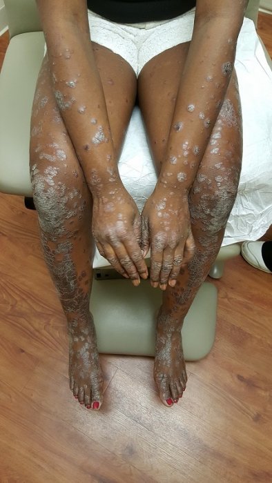
The patient was referred to allergist for testing. She was found to be significantly atopic with positive reactions to ragweed, weeds, grass, dust mite, and cats. Her elevated serum tryptase also indicated systemic involvement, which prompted a bone marrow biopsy. The patient’s bone marrow biopsy further confirmed the diagnosis of systemic mastocytosis. The patient has no GI symptoms, bone pain, diarrhea, etc. She is taking fexofenadine in the morning and cetirizine in the evening. She also has an epinephrine pen and was instructed to be cautious with alcohol, NSAIDs, aspirin, bee stings, local trauma to the skin lesions, etc.
REFERENCES:
Soter, N. A. (1991). The Skin in Mastocytosis. J Invest Dermatol, 96(3), 32S-39S. PMID: 2002678
Vano-Galvan, S., De la Hoz, B., Núñez, R., & Iaen, P. (2010). Indolent Systemic Mastocytosis. IMAJ, 12, 185-188. PMID: 20345674
Johnson, M. R., Verstovsek, S., Jorgensen, J. L., Manshouri, T., Luthra, R., et al. (2009). Utility of the World Health Organization Classification Criteria for the Diagnosis of Systemic Mastocytosis in Bone Marrow. Mod Pathol, 22, 50-57. doi:10.1038/modpathol.2008.184. PMID: 18806881
Metcalfe, D. D. (2003). The Mastocytosis Syndrome. In: Fitzpatrick’s Dermatology in General Medicine. New York (NY): McGraw-Hill; 2003. p. 1603-1608.
Kennedy, R. J., Scoffield, J. L., & Garstin, W. I. H. (1999). An Unusual Presentation of Systemic Mastocytosis. J Clin Pathol, 52, 301-302. doi:10.1136/jcp.52.4.301. PMID: 10440165
Lanternier, F., Cohen-Akenine, A., Palmerini, F., Feger, F., Yang, Y., et al. (2008). Phenotypic and Genotypic Characteristics of Mastocytosis According to the Age of Onset. PLoS ONE, 3(4), e2058. doi:10.1371/journal.pone.0002058. PMID: 18446280
Akin, C., & Metcalfe, D. D. (2004). Systemic Mastocytosis. Annu Rev Med, 55, 419-432. doi:10.1146/annurev.med.55.090102.103030. PMID: 14745316
Lim, K., Tefferi, A., Lasho, T. L., Finke, C., Patnaik, M., et al. (2009). Systemic Mastocytosis in 342 Consecutive Adults: Survival Studies and Prognostic Factors. Blood, 113(23), 5727-5736. doi:10.1182/blood-2008-12-196152. PMID: 19372337
Inamadar, A. C., & Palit, A. (2006). Cutaneous Mastocytosis: Report of Six Cases. Indian J Dermatol Venereol Leprol, 72(1), 50-53. PMID: 16564979
Briley, L. D., & Phillips, C. M. (2008). Cutaneous Mastocytosis: A Review Focusing on the Pediatric Population. Clin Pediatr (Phila), 47(8), 757-761. doi:10.1177/0009922808314966. PMID: 18625749
Peterson, A. H. (1984). Systemic Mastocytosis: Case Report and Literature Review. J Natl Med Assoc, 76(5), 469-473. PMID: 6379044
Metcalfe, D. D. (1991). Classification and Diagnosis of Mastocytosis: Current Status. J Invest Dermatol, 96(3), 2S-4S. PMID: 2002674
Bains, S. N., & Hsieh, F. H. (2010). Current Approaches to the Diagnosis and Treatment of Systemic Mastocytosis. Ann Allergy Asthma Immunol, 104, 1-10. doi:10.1016/j.anai.2010.09.005. PMID: 20923212
Stevens, E. C., & Rosenthal, N. S. (2001). Bone Marrow Mast Cell Morphologic Features and Hematopoietic Dyspoiesis in Systemic Mast Cell Disease. Am J Clin Pathol, 116, 177-182. doi:10.1309/7X1G-MTT8-3Q9G-UX77. PMID: 11474596


