Presenter: Kate Kleydman, DO
Dermatology Program: Saint. Barnabas Hospital
Program Director: Cindy Hoffman, DO FAOCD
Submitted on: November 14, 2011
CHIEF COMPLAINT: “My skin is so stiff”
CLINICAL HISTORY: A 52-year-old African-American female presented with complaints of having “stiff skin” that progressively impaired her movement over the past five years. The skin “tightness” had started on the body, and then progressed to include her hands, trunk, legs, and finally face. She complained of constant pain, with restrictions of movement requiring the use of a walker. She experienced worsening of the pain in her hands, accompanied by color changes and tingling in cold weather. Her review of systems was positive for difficulty swallowing, acid reflux, dyspnea on exertion, nonproductive cough, diffuse arthralgias and myalgias, subjective decreased range of motion, and chronic fatigue. No previous treatment. Her past medical history was significant for hypertension and gastroesophageal reflux disease. The patient was taking Lisinopril and Percocet and denied alcohol and drug use. Her family history was negative for any significant dermatologic diseases or autoimmune disorders.
PHYSICAL EXAM:
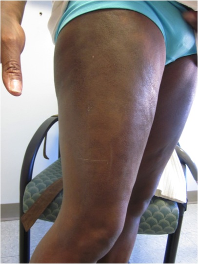
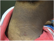
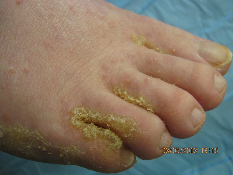
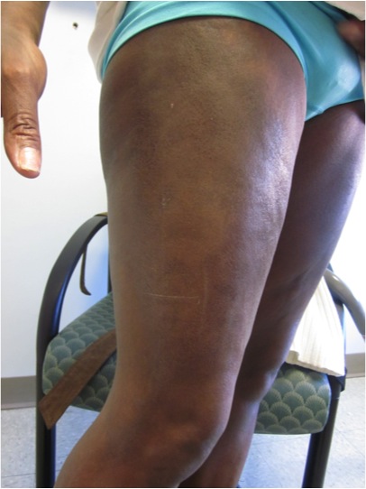
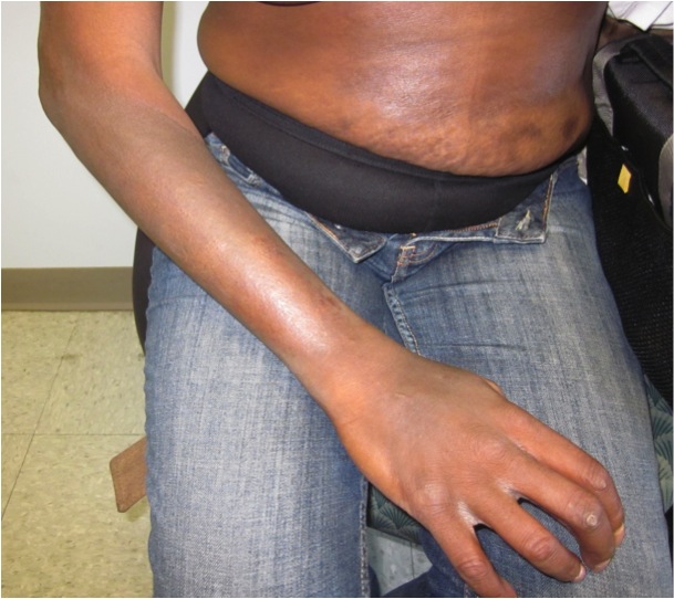
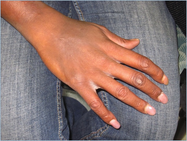
Physical exam revealed taught, shiny skin with a salt and pepper pigmentation throughout the abdomen, back, and extremities. There was complete hair loss of both upper and lower extremities. Hyperpigmented thickened plaques spanned the trunk, thighs, and upper extremities bilaterally. There were patchy areas of sparing in affected locations, including complete sparing of both antecubital fossae. The hands and feet revealed sclerodactyly. There were ulcerations on the dorsal aspect of the skin overlying the proximal interphalangeal (PIP) joints of the right second and third digits.
LABORATORY TESTS:
ANA – high positive, 1:320, nucleolar pattern
GFR – 51.98
UA – WNL
CMP – WNL
RNP ab – negative
Centromere ab – negative
SCL 70 ab – negative
RF – negative
CBC – Hg 11.0, Hct 33.9, MCV 76.2
DERMATOHISTOPATHOLOGY:
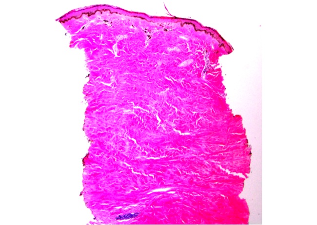
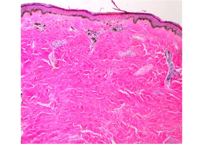
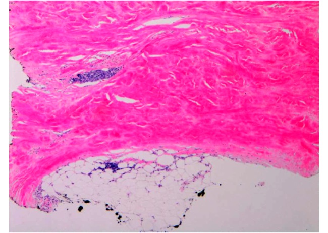
A 3 mm punch biopsy was taken on the left upper arm, revealing dermal pallor and thick, closely packed, hyalinized collagen bundles in the lower dermis. There is a loss of adventitial fat resulting in “trapped” eccrine glands with a sparse deep lymphoplasmacytic infiltrate. There is extensive pan-dermal fibrosis and collagen noted extending to subcutis.
DIFFERENTIAL DIAGNOSIS:
1. Scleredema
2. Scleromyxedema
3. Eosinophilic Fasciitis
4. Eosinophilia Myalgia Syndrome
5. Diabetic Stiff Skin

