CORRECT DIAGNOSIS:
Lichen Amyloidosis
DISCUSSION:
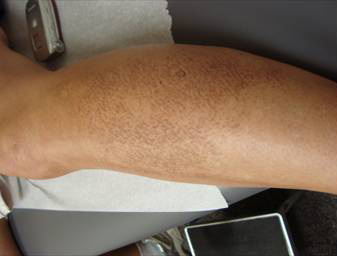
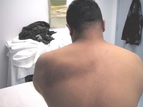
Lichen amyloidosis is the most common form of primary cutaneous amyloidosis. Lichen amyloidosis presents as intensely pruritic, red-brown hyperkeratotic papules most commonly seen on the pretibial surfaces. Usually, it presents as unilateral lesions with the eventual symmetrical distribution. Friction or chronic rubbing of the skin contributes to the cutaneous findings. Asian, Hispanic, or Middle Eastern individuals appear to be predisposed to this condition. Lichen amyloidosis is more common in men and persons aged 50-60 years old.
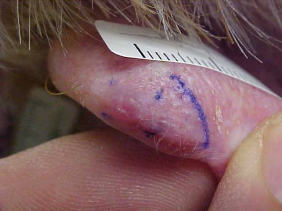
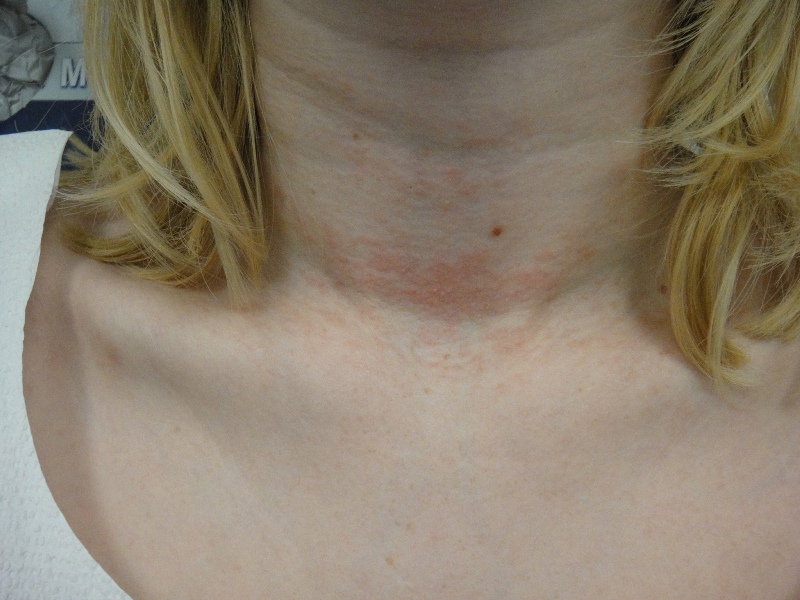
There are three primary forms of cutaneous amyloidosis: lichen amyloidosis, macular amyloidosis, and nodular amyloidosis. Dermatologists are more likely to encounter the cutaneous forms of amyloid, which have a benign course, and less frequently the systemic form of amyloid, which has fewer cutaneous findings. Systemic amyloidosis results from amyloid protein deposition in organ systems and is associated with significant morbidity and a high mortality rate.
Histopathologically, amyloid deposits are found in the papillary dermis, usually at the tips of the dermal papillae. Amyloid appears as an amorphous, eosinophilic, fissured substance. Lichen amyloidosis is distinguished from other cutaneous forms by the presence of marked epidermal changes including hyperkeratosis and acanthosis. Several stains demonstrate the presence of amyloid deposits in the skin. The best-known stain is Congo red, which under polarizing light demonstrates apple-green birefringence. Other stains include periodic acid-Schiff (PAS), methyl violet, crystal violet, various cotton dyes (eg, pagoda red, Sirius red) and the fluorescent dyes, thioflavin-T and Phorwhite BBU.
TREATMENT:
The patient was treated with 30% topical urea to the affected areas once daily, which greatly reduced the thickness of the lesions and decreased pruritus.
Since chronic rubbing seems to be the inciting factor in the deposition of amyloid in lichen amyloidosis, treatment requires identification of the underlying cause of pruritus, habit, or neuropathy. Treatment modalities include oral sedating antihistamines, topical menthol, topical and intralesional steroids, urea agents, and narrow-band UVB phototherapy. Surgical strategies including microdermabrasion and laser ablation have been used, however, the pruritus and lesions themselves have been reported to recur after these treatments.
REFERENCES:
Wang, W. J. (1990). Clinical features of cutaneous amyloidoses. Clinical Dermatology, 8, 13-19. PMID: 2191793
Weyers, W., et al. (1997). Lichen amyloidosus: A consequence of scratching. Journal of the American Academy of Dermatology, 37(6), 923-928. doi:10.1016/S0190-9622(97)70041-9. PMID: 9408425
Touart, D. M., & Sau, P. (1998). Cutaneous deposition diseases. Part I. Journal of the American Academy of Dermatology, 39(2 Pt 1), 149-171. doi:10.1016/S0190-9622(98)70066-4. PMID: 9700182
Lambert, W. C. (1990). Cutaneous deposition disorders. In E. R. Farmer & A. F. Hood (Eds.), Pathology of the Skin (Vol. 432, p. 50). Norwalk, Conn: Appleton & Lange.


