CORRECT DIAGNOSIS:
Phytophotodermatitis
DISCUSSION:
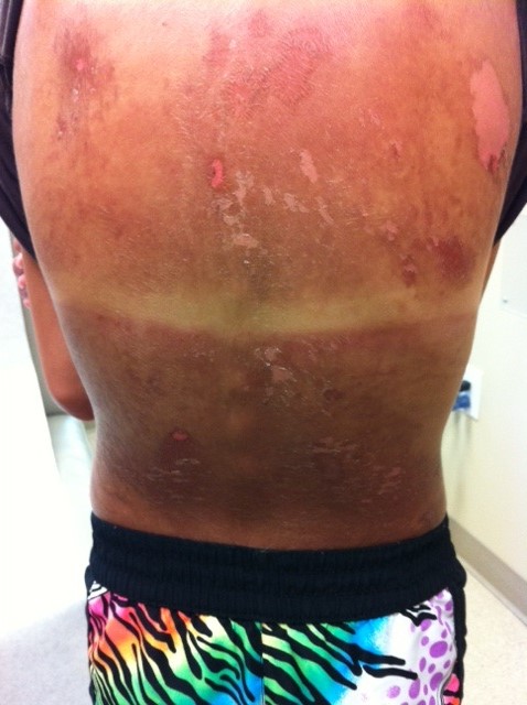
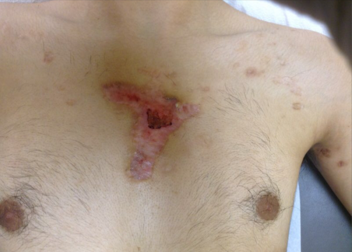
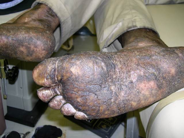
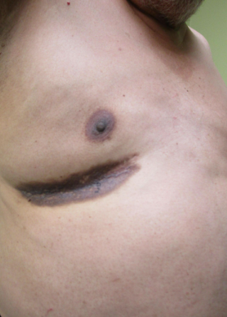
Phytophotodermatitis, also known as Berloque Dermatitis, is a cutaneous phototoxic eruption as a result of contact with furocoumarin substances and long-wave ultraviolet (UVA 320-380 nm) radiation. The cutaneous lesions typically appear approximately 24 hours after exposure, and peaks at 48 to 72 hours. In the acute phase, the affected patches initially range from erythematous macules, patches, plaques, vesicles, and or bullae, with tender areas similar to a severe sunburn. In severe cases, systemic symptoms including fever, nausea, and vomiting may occur. Post-inflammatory hyperpigmentation, characteristically grey-brown in presentation, may ensue following the acute lesions. The hyperpigmentation may, however, take several weeks to months to completely resolve after cessation of the offending substance.
Bergamot oil is one of the originally described etiologies for this condition, and the oil of Bergamot containing 5-methoxypsoralen found in many commercial products is restricted. However, other psoralens or furocoumarins, such as lemons, limes, figs, parsley, and celery also contain psoralens and are well documented to have phototoxic reactions. Other plants associated with phytophotodermatitis include carrots, fennel, dill, buttercup, and mustard. , Limes have a high concentration of psoralens and are often a highly reported culprit of phytophotodermatitis. Additionally, there have been reports of ingestion of psoralen rich foods, such as celery may cause generalized phototoxicity. The long wave UVA radiation acts on bergapten, a photoactive psoralen component of bergamot oil, resulting in erythema, hyperpigmentation, and in severe cases bullous reactions in the areas exposed. There is documentation that in ancient times, the principles of phototoxic reactions were used as therapy for induction of hyperpigmentation in syndromes such as vitiligo, such as the use of bavachee seeds in India as early as 1400 BC.
Phytophotodermatitis may clinically present similar to contact dermatitis, chemical burns, and has been mistaken in pediatric patients for child abuse. Other differential diagnosis includes herpes zoster, porphyria cutanea tarda, and thermal burns. Clinical history is important in confirming the diagnosis. The cutaneous findings are commonly unusual, asymmetric patterns in sun-exposed areas with recent exposure to a photosensitizing plant and areas of post-inflammatory hyperpigmentation. As the lesions appear in areas exposed to the citrus fruits in combination with sun exposure, hyperpigmented linear streaks may be one form of clinical presentation. The hands and mouth may be a commonly involved location as well.
Management in phytophotodermatitis is primarily to stop the offending agent to prevent future recurrences. Further treatment is predominately directed towards symptomatic relief, specifically in diminishing the inflammatory response, which includes cool wet dressings, topical corticosteroids, antipruritic agents, soothing lotions, and potentially systemic corticosteroids if severe or lesions are too extensive. Patients must be encouraged to use sunscreen to prevent further or chronic hyperpigmentation.8 Bleaching agents, such as hydroquinone can be used if hyperpigmentation persists and is bothersome to the patient.
TREATMENT:
Our patient was treated with topical corticosteroids and moisturizers and improved in the subsequent weeks with sun avoidance and sunscreen.
REFERENCES:
Smith, E., Kiss, F., Porter, R. M., & Anstey, A. V. (2011). A review of UVA-mediated photosensitivity disorders. Photochemical & Photobiological Sciences, 11(1), 199-206. doi:10.1039/C0PP00046A
MacFarlane, D. F., & DeLeo, V. A. (1995). Phototoxic and photoallergic dermatitis. In J. D. Guin (Ed.), Practical contact dermatitis (pp. 83-92). New York, NY: McGraw-Hill Co.
Pathak, M. A. (1986). Phytophotodermatitis. Clinical Dermatology, 4, 102-121.
Gould, J. W., Mercurio, M. G., & Elmets, C. A. (1995). Cutaneous photosensitivity diseases induced by exogenous agents. Journal of the American Academy of Dermatology, 33, 551-573. doi:10.1016/S0190-9622(95)90266-1
Lovell, C. R. (Ed.). (1993). Phytophototoxic reactions. In Plants and the skin (pp. 64-95). Boston: Blackwell Scientific.
Wagner, A. M., Wu, J. J., Hansen, R. C., et al. (2002). Bullous phytophotodermatitis associated with high natural concentrations of furocoumarins in limes. American Journal of Contact Dermatitis, 13, 10-14. doi:10.1056/NEJMoa1003014
Lovell, C. R. (1997). Phytodermatitis. Clinical Dermatology, 15, 607-613.
Marzulli, F. N., & Maibach, H. I. (1970). Perfume phototoxicity. Journal of the Society of Cosmetic Chemists, 21, 695-715.
Solis, R. R., Dotson, D. A., & Trizna, Z. (2000). Phytophotodermatitis: A sometimes difficult diagnosis. Archives of Family Medicine, 9, 1195-1196. doi:10.1001/archfami.9.12.1195
Juckett, G. (1996). Plant dermatitis: Possible culprits go far beyond poison ivy. Postgraduate Medicine, 100, 159-171. doi:10.3810/pgm.1996.09.1009
Weber, I. C., Davis, C. P., & Greeson, D. M. (1999). Phytophotodermatitis: The other “lime” disease. Journal of Emergency Medicine, 17, 235-237. doi:10.1016/S0736-4679(99)00108-3


