Presenter: Katherine Johnson, D.O.
Dermatology Program: Botsford Hospital
Program Director: Annette LaCasse, D.O.
Submitted on: June 2, 2012
CHIEF COMPLAINT: Lower extremity swelling, pruritis, and pain
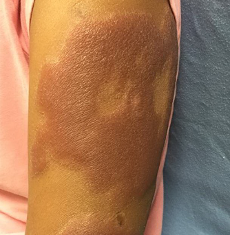
CLINICAL HISTORY: A 67-year-old Caucasian male presented to the clinic with a chief complaint of lower extremity swelling, pruritus, and pain. He also complained of discoloration of his lower extremities, right arm, and axillae, stating that one of his feet was “turning black”. Over a one-month duration, the patient noticed dark patches developing on his lower extremities, right upper extremity, and axillae. There was associated edema in his lower extremities accompanied by pain and pruritus. The patient visited urgent care, an emergency room, and his podiatrist for the chief complaint. Laboratory studies, radiographs, and an EKG were all found to be normal. The patient’s past medical history included renal transplantation in October 2010, and was maintained on mycophenolate mofetil 1g PO BID, tacrolimus 3mg PO BID, and prednisone 10 mg PO daily. Pertinent family medical history included a brother who was deceased due to malignant melanoma found in the axillae.
PHYSICAL EXAM:
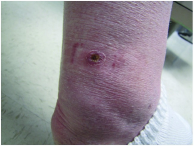
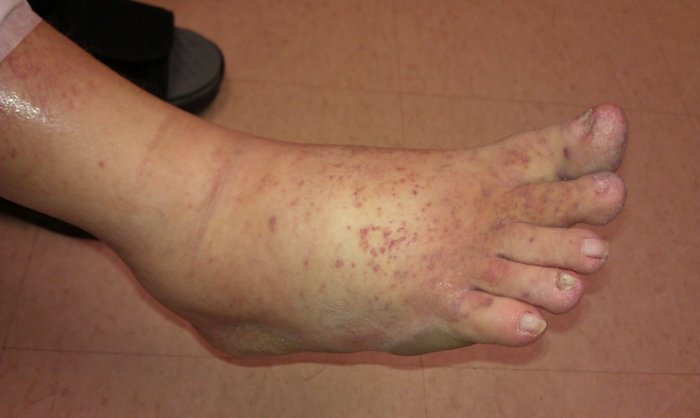
Physical examination revealed violaceous papules and plaques over the extensor surfaces of the right upper and lower extremities. In addition, there were violaceous plaques on the bilateral plantar surfaces and 2-4 mm erythematous macules on the bilateral dorsal feet and anterior lower legs. The right lower extremity had pitting edema and pain upon palpation.

LABORATORY TESTS:
An HIV screen was negative.
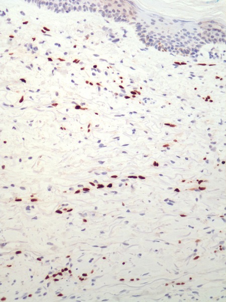
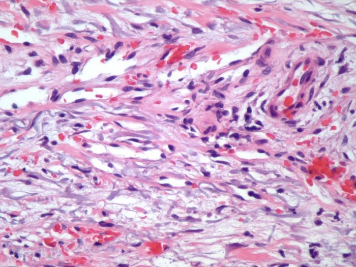
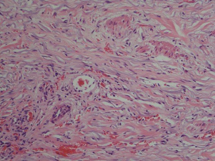
DERMATOHISTOPATHOLOGY:
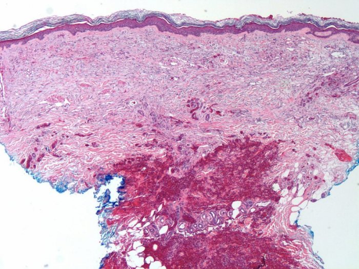
A punch biopsy of the right upper extremity and right plantar foot was performed, which demonstrated proliferation of endothelial cells forming bizarre-shaped, thin-walled vessels that followed pre-existing vascular plexuses. A CD31 stain was performed, highlighting the vascular proliferative changes and an HHV-8 stain decorated with many spindle cells that were associated with the vessels.
DIFFERENTIAL DIAGNOSIS:
1. Kaposi’s Sarcoma
2. Glomuvenous malformation
3. Angiosarcoma
4. Spindle cell hemangioma
5. Metastatic malignant melanoma