CORRECT DIAGNOSIS:
Metastatic Breast Adenocarcinoma
DISCUSSION:
Cutaneous metastases are of diagnostic importance because they may be the first manifestation of an undiscovered internal malignancy (as was the case with our patient) or the first indication of metastasis of a supposedly adequately treated malignancy. The site of the primary tumor, if unknown, may be suspected by the histopathology and immunoperoxidase studies.
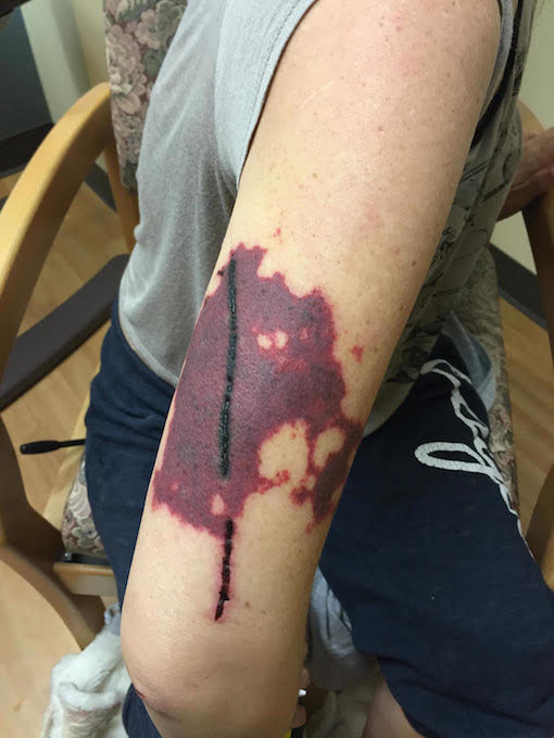
Cutaneous metastases of breast carcinoma that occur predominantly by lymphatic dissemination include inflammatory carcinoma, carcinoma en cuirasse, telangiectatic and nodular carcinoma, and carcinoma of the inframammary crease. Alopecia neoplastica and mammary carcinoma of the eyelid are probably caused by hematogenous spread.
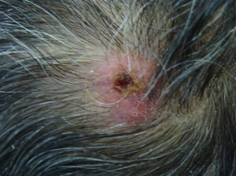
Alopecia neoplastica occurs as oval plaques or patches of the scalp and may be confused clinically with alopecia areata or scarring alopecia. Alopecia neoplastica is defined as hair loss secondary to a visceral malignancy that has metastasized to the scalp. Although cancers that primarily present in the skin-such as basal cell carcinoma, squamous cell carcinoma, and angiosarcoma can morphologically appear as alopecia, this presentation does not fulfill the definition of alopecia neoplastica.
The tumor cells have elongated nuclei similar to fibroblasts, but the nuclei are larger, more angular, and more deeply basophilic. The tumor cells often lie singly; however, in some areas, they may form small groups or single rows between fibrotic and thickened collagen bundles. This latter feature of “Indian filing” is of particular diagnostic importance and may be seen in any histologic variety of metastatic breast cancer.
Immunoperoxidase Studies; Cutaneous metastases are positive with most cytokeratins, except for CK20, and often are positive with epithelial membrane antigen (EMA) and carcinoembryonic antigen (CEA). Expression of CK7 was found in the majority of cases of carcinoma, with the exception of those arising from the colon, prostate, kidney, thymus, carcinoid tumors of the lung and gastrointestinal tract, and Merkel cell tumors of the skin.
Ironically, the lack of distinctive clinical features may be a clue to the diagnosis of cutaneous metastases. Anatomic location, the primary morphology, and lesional color can sometimes be helpful. When distinct clinical features are not present, the primary morphology of most metastases is a papule or especially a nodule. Most sources cite the usual morphologic differential diagnosis of a dermal/subcutaneous nodule, i.e. a cyst, lipoma, appendageal tumor, or fibroma. Since the pathogenesis of metastasis intimately involves angiogenesis, the primary color is often some shade of red. As a result, metastatic carcinoma can simulate a number of benign and malignant vascular lesions.
TREATMENT:
Therapy can be tailored to treat any combination of primary cancer and cutaneous metastasis. Treatment of the latter includes chemotherapy, immunotherapy, radiotherapy, excision, heat, and observation. Occasionally intralesional and topical (e.g. imiquimod) agents are employed. Tumor burden, maintaining the daily function, cosmesis, and the possibility of pain, bleeding, and infection are factored into the treatment algorithm. Although the disease course depends on the underlying malignancy and its therapeutic response, cutaneous metastasis is a marker of advanced disease, and, historically, life expectancy has been short.
For this patient, an ultrasound-guided needle core biopsy of the left breast revealed an invasive ductal carcinoma. A Brain MRI revealed small right temporal lobe metastasis. The Patient was admitted to hospice and expired 3 months later.
REFERENCES:
Schwartz, R. A. (1995). Cutaneous metastatic disease. Journal of the American Academy of Dermatology, 33, 161. https://doi.org/10.1016/S0190-9622(95)90169-6 PMID: 7610384
Wesche, W. A., Khare, V. K., Chesney, T. M., et al. (2000). Non-hematopoietic cutaneous metastases in children and adolescents: Thirty years experience at St. Jude Children’s Research Hospital. Journal of Cutaneous Pathology, 27, 485–492. https://doi.org/10.1034/j.1600-0560.2000.270906.x
Reingold, I. M. (1966). Cutaneous metastases from internal carcinoma. Cancer, 19, 162-168. https://doi.org/10.1002/1097-0142(196601)19:1<162::AID-CNCR2820190118>3.0.CO;2-K
Brownstein, M. H., & Helwig, E. B. (1973). Spread of tumors to the skin. Archives of Dermatology, 107, 80-86. https://doi.org/10.1001/archderm.1973.01620010084012 PMID: 4719744
Murray, S., Simmons, I., & James, C. (2003). Cutaneous angiosarcoma of the face and scalp presenting as alopecia. Australasian Journal of Dermatology, 44, 273-276. https://doi.org/10.1111/j.1440-0960.2003.00609.x


