Presenter: Jared Heaton D.O., Brooke Walls D.O., Julian M. Ngo D.O.
Dermatology Program: NSUCOM/Largo Medical Center
Program Director: Richard Miller D.O.
Submitted on: October 6, 2012
CHIEF COMPLAINT: Recurrent malodorous infection of the scalp
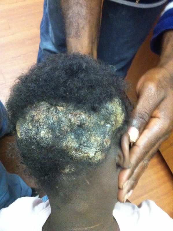
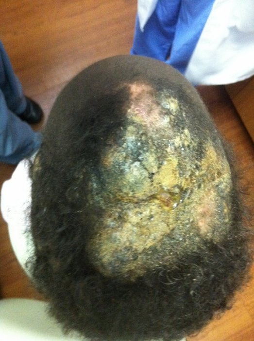
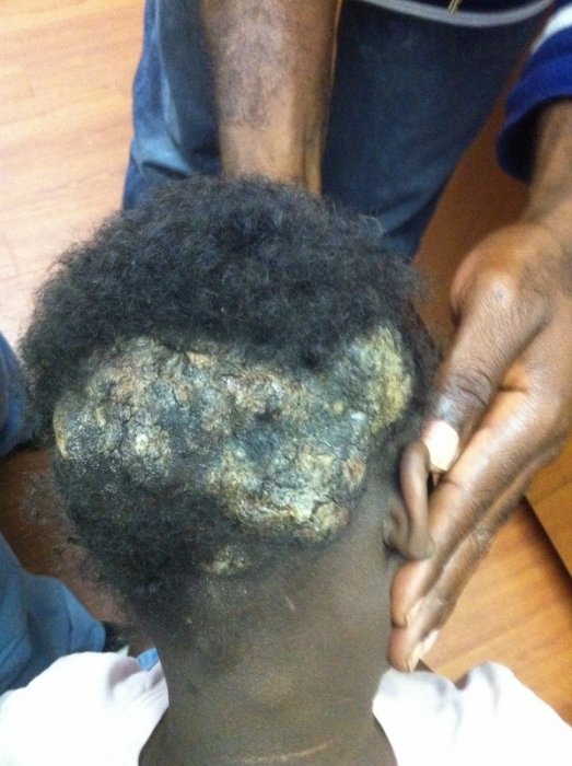
CLINICAL HISTORY: A 5 y/o AA female presented for evaluation of recurrent malodorous infection of the scalp. This patient has a history of recurrent bacterial and fungal infections of the scalp for the past two to three years. She was recently admitted to All Children’s Hospital for pyoderma of the scalp secondary to MRSA where she was treated with IV antibiotics and surgical debridement. She had a previous surgical debridement of the scalp 1 year ago for the same condition recalcitrant to oral and topical medications. Several weeks after her most recent discharge from the hospital, the patient again developed a large fungating verrucous plaque of the scalp. Surgical debridement of the scalp secondary to pyoderma was performed in June 2012 and April 2011.
Medications: Griseofulvin, Fluconazole, Cephalexin, Prednisone, Ammonium Lactate, Calcipotriene cream, Mupirocin ointment, BPO 5% wash, Topical Steroids, Prednisolone ophthalmic suspension.
Other information: Mother and Father are both deaf. The patient’s brother is also deaf and has similar skin findings. The patient also has a history of severe asthma, allergies, early cataracts, and bilateral conjunctivitis. She has had multiple positive bacterial cultures from the scalp (MRSA) in the past.
PHYSICAL EXAM:
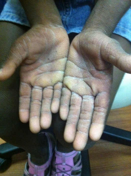
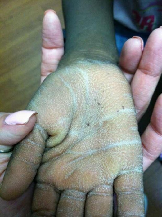
A large hyperkeratotic fungating plaque involving the frontal, parietal and occipital scalp with overlying thick yellow/white stuck-on scale. Some purulent discharge of the plaque was noted. Exam of the patient’s hands and feet revealed palmar-plantar hyperkeratosis with a stippled appearance. Visible posterior cervical lymphadenopathy was also appreciated.
LABORATORY TESTS:
CBC: WBC 7.65 with 60% lymphocytes and 2.6% neutrophils. IgG 1740 (wnl), IGA 271 (mildly elevated), IgM 138 (wnl). Tissue culture negative for fungal elements
DERMATOHISTOPATHOLOGY:
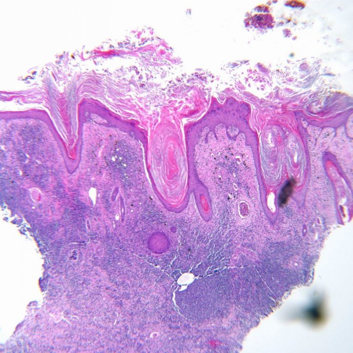
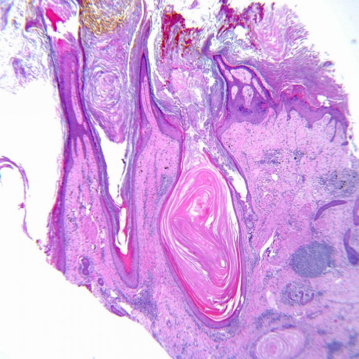
Marked epidermal hyperplasia and hyperkeratosis, with cystically dilated and ruptured follicular infundibula, dense mixed dermal inflammation, and superficial impetiginization. There is superficial colonization with gram-positive bacterial cocci, which may be contributing to the inflammatory process resulting in disruption of follicular epithelium. Courtesy of Gulf Coast Pathology: Michael Heaphy, MD
DIFFERENTIAL DIAGNOSIS:
1. Wiskkot-Aldrich syndrome
2. KID Syndrome
3. Folliculitis Decalvans
4. IFAP (Ichthyosis, Follicularis with Alopecia and Photophobia) syndrome
5. Vohwinkel syndrome

