Presenter: Michael Centilli, DO
Dermatology Program: Botsford/McLaren Oakland
Program Director: Annette LaCasse, DO
Submitted on: January 30, 2013
CHIEF COMPLAINT: Generalized eruption of hyperkeratotic, verrucous papules
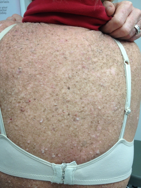
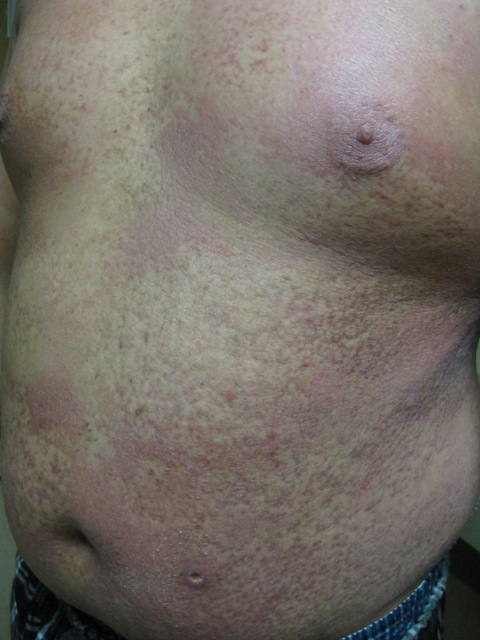
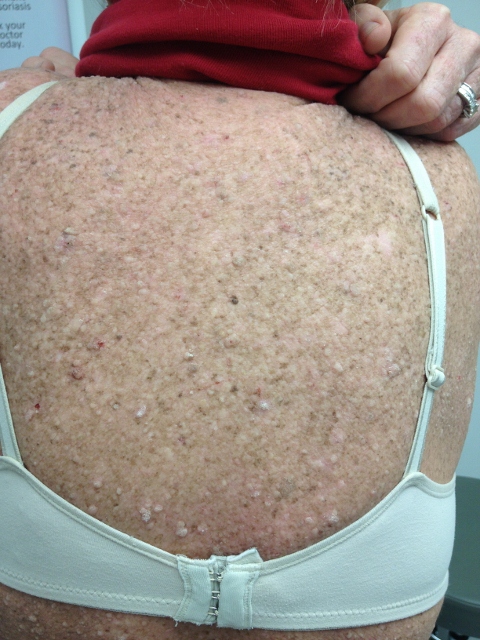
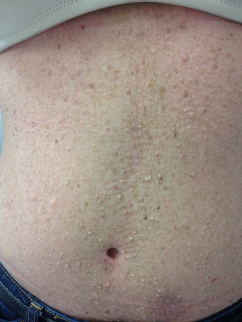
CLINICAL HISTORY: 57-year-old Caucasian female presented with a chief complaint of an 8-month history of a generalized eruption of hyperkeratotic, verrucous papules beginning in her left axilla with subsequent spread to her back, trunk, face, and all four extremities. Review of systems was positive for extensive pruritis and mild dysphagia with solid food. Prior to our consultation the patient had tried multiple therapies administered by several dermatologists, including cryotherapy, intralesional injection of 0.1 cc of candida antigen, the patient did not return for serial injections, and topical salicylic acid resulting in second-degree burns. Past medical history was positive for basal cell carcinoma of the left lower thigh, an abnormal pap smear of undetermined significance, a benign mass of the right breast, and breast augmentation. HIV testing had been performed in the past year and was negative.
PHYSICAL EXAM:
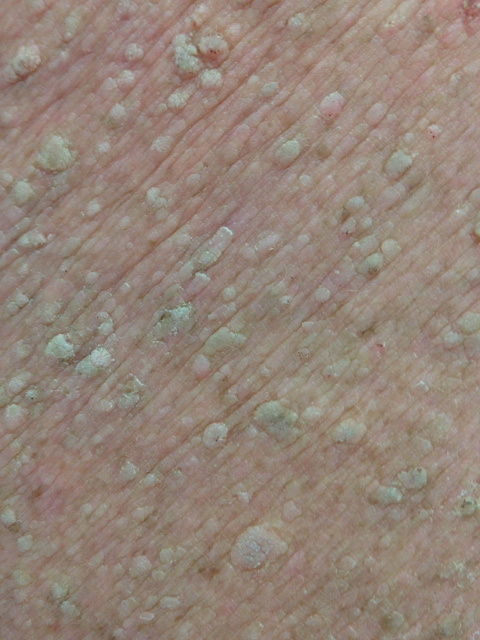
Examination revealed numerous flesh-colored to tan verrucous papules that covered approximately 90% of her body surface area. Well-demarcated second-degree burns were also present on the right thigh and bilateral upper arms. The palmar surface of each hand was hyperkeratotic with a coarse texture.

LABORATORY TESTS:
Endoscopy (with esophageal biopsy) revealed a large, friable, polypoid lesion located at the distal esophagus and gastroesophageal junction. The biopsy of the mass was consistent with moderate to poorly differentiated adenocarcinoma. CT of the thorax with IV contrast (with CT guided needle biopsy) showed a 2.1 cm right hepatic lobe mass. A biopsy was consistent with metastatic adenocarcinoma.
DERMATOHISTOPATHOLOGY:
Two prior biopsies by different dermatologists were read as verruca vulgaris and seborrheic keratosis with verrucous changes. A repeat 4-mm punch biopsy taken from the back was consistent with verruca vulgaris, and a shave biopsy taken from the thigh was consistent with seborrheic keratosis with verrucous changes. No evidence of epidermodysplasia verruciformis was seen. HPV typing of the lesions was also negative.
DIFFERENTIAL DIAGNOSIS:
1. Epidermodysplasia Verruciformis
2. Paraneoplastic Syndrome- Sign of Leser-Trelat
3. Verruca Vulgaris (Disseminated secondary to immunocompromise)
4. Verrucous Seborrheic Keratoses

