Presenter: Peter Knabel DO, Cathy Koger DO, Stephen Plumb DO, Chris Cook DO
Dermatology Program: Northeast Regional Medical Center
Program Director: Dr. Lloyd Cleaver
Submitted on: March 1, 2013
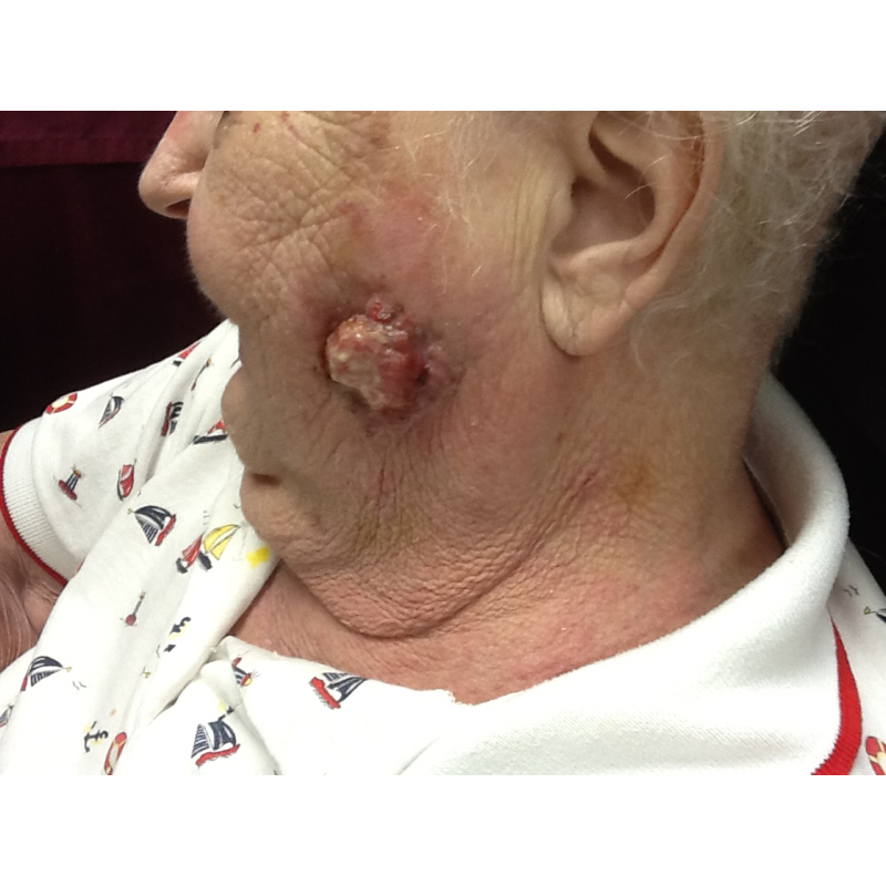
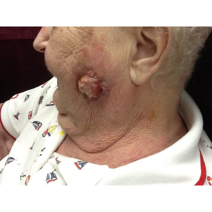
CHIEF COMPLAINT: Rapidly expanding, exophytic, painful ulceration on her left cheek
CLINICAL HISTORY: The patient is an 85-year-old Caucasian female who presented with a rapidly expanding, exophytic, painful ulceration on her left cheek. The lesion is a friable, tender, erythematous nodule that had rapidly grown in the previous 6 months. No previous treatment. The patient resides in an assisted living facility, and they had provided symptomatic relief for her as needed. The patient has a past medical history consistent with sick sinus syndrome, rheumatoid arthritis, mild depression, and hypertension. She has no personal history of skin cancer or skin disorders. Her family history included a father that died of pancreatic cancer and a brother of unknown malignancy. She has never smoked or abused alcohol and has no known risk factors for sexually transmitted diseases.
PHYSICAL EXAM:
The patient presented with a 3.0 x 2.7cm nonpigmented, ulcerated, exophytic plaque on the left malar area. Patient also had some erythematous scaly plaques on her arms and trunk as well as some hyperkeratotic plaques on her hands. She had no cervical, axial, or inguinal lymphadenopathy.
LABORATORY TESTS:
No laboratory tests were ordered upon the patient’s initial presentation.
DERMATOHISTOPATHOLOGY:
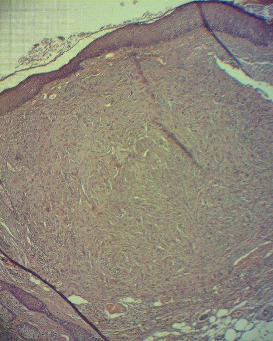
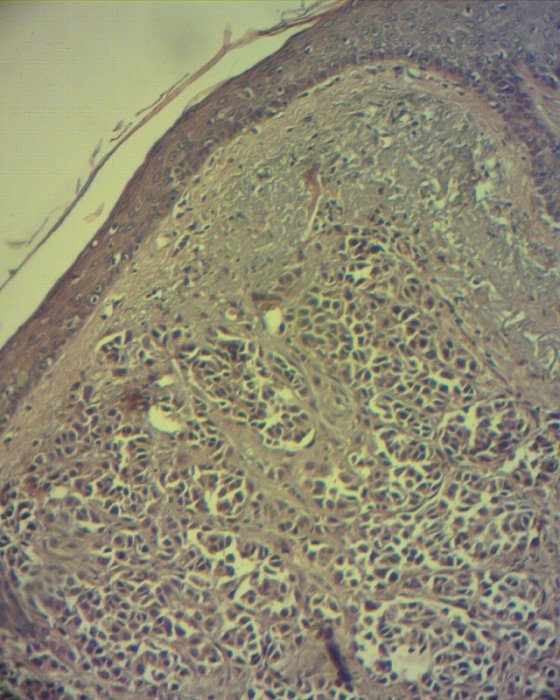
Histology reveals sections with ulceration as well as a cellular proliferation of oval to spindled cells in the superficial dermis. In multiple areas, the cells had a nested appearance with numerous mitotic figures.
DIFFERENTIAL DIAGNOSIS:
1. Basal Cell Carcinoma
2. Melanoma
3. Atypical Fibroxanthoma
4. Merkel Cell Carcinoma
5. Squamous Cell Carcinoma

