CORRECT DIAGNOSIS:
Melanoma with multiple histologic subtypes
DISCUSSION:
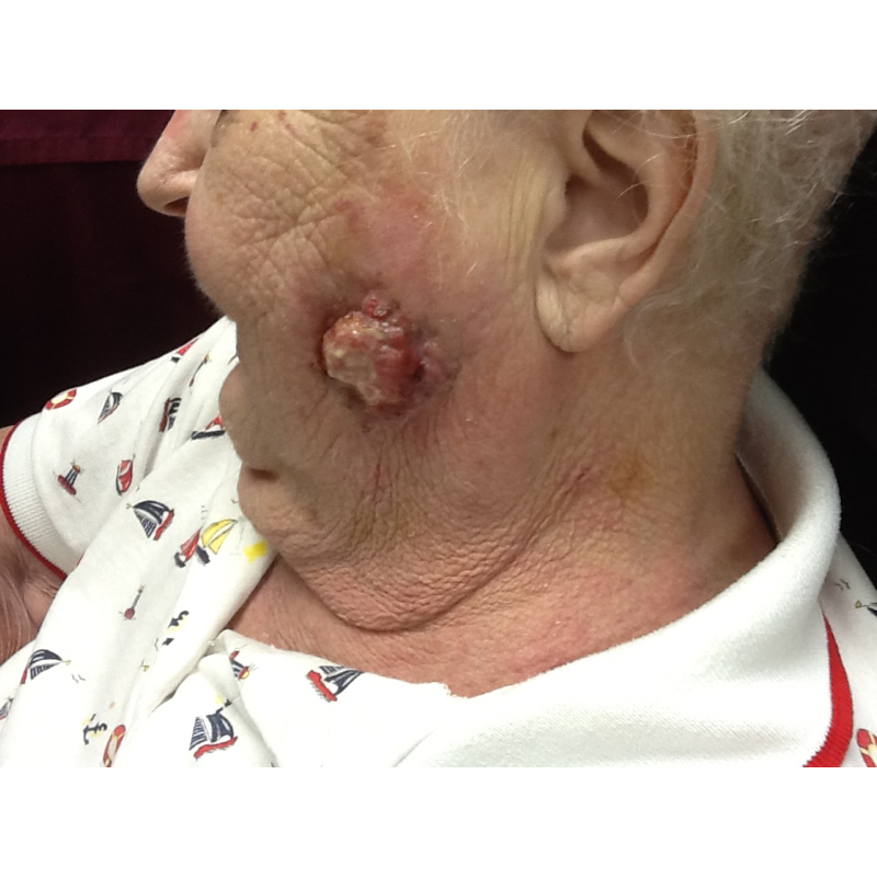
Excision was performed 3 weeks later on this lesion (which had expanded to 4.8 x 4.2cm) that revealed melanoma with multiple histologic subtypes, including nodular, superficial spreading, spindled, amelanotic, and desmoplastic. Histologic features of the excision included a Breslow depth of 8mm, broad ulceration, high mitotic rate, and peri-vascular with possible peri-neural invasion.
There is a paucity of literature regarding melanoma with multiple histologic subtypes. It is possible an underreported entity or just extremely rare. Multiple histologic subtype melanoma may have contributed to the rapid appearance and progression of this patient’s lesion, but again there are no other literature reports to support this conclusion.
Interestingly this patient had a father who died of pancreatic cancer and a brother of unknown malignancy. As has been elucidated in research, there are many pathways that have been implicated in the development of melanoma some of which include RAS/MAPK, c-KIT, and Rb. The RAS/MAPK pathway has been associated with the BRAF/V600E mutations, where melanomas tend to appear in younger individuals and have been shown to respond to Zelboraf/vemurafenib. The c-KIT pathway of melanoma development is typically site-specific, with a predilection for mucosal, acral, and chronically sun-damaged skin. These lesions have been shown to respond to Gleevec/imatinib as well as Tasigna/nilotinib. Finally, the Rb pathway has been associated with CDKN2A or CDK4 mutations which seem to have a familial inheritance pattern. This pathway has also been shown to be associated with increased risk for pancreatic cancer. Perhaps our patient inherited a gene mutation from her father that predisposed her to this aggressive melanoma, which ultimately led to her mortality. Hopefully, in the future genetic testing will help identify patients like her so we can be offer better preventative care.
In short, our 85-year-old female presented to us with a very unusual and aggressive form of melanoma that revealed multiple histologic subtypes and a poor prognosis. There are very few other cases in the literature with even two histologic subtypes, making this case unique and unusual. Sadly our patient passed away within six months of initial presentation and her tumor had continued to progress. Given her family history of pancreatic cancer and her subsequent melanoma, it is possible that she may have a CDK4 gene mutation that predisposed her to this tumor.
TREATMENT:
Our patient had a recurrence of this melanoma, even after her excision. An MRI of the brain, as well as a chest x-ray, didn’t reveal any distant metastases, but the patient was referred to oncology for other possible treatment options. Radiation was deemed the most appropriate treatment given her age, the tumor type, and her comorbidities. The patient and her family deferred this treatment, so she was placed on hospice and sadly passed away a few months later.
REFERENCES:
Bolognia, J. L., Jorizzo, J. L., & Rapini, R. P. (2003). Dermatology (2nd ed., Vol. 2, pp. 1745-1760). Elsevier.
James, W. D., Berger, T. G., & Elston, D. M. (2006). Andrews’ Diseases of the Skin (11th ed., pp. 680-690). Saunders.
Rapini, R. P. (2009). Practical Dermatopathology (1st ed., pp. 273-279). Elsevier.
Rojas, C. A., & Ahn, C. S. (2009). The evolving role of the dermatopathologist in hematology/oncology. Hematology/Oncology Clinics of North America, 23(3), 501-513. https://doi.org/10.1016/j.hoc.2009.04.001
Goldfarb, D. M., & Jellinek, N. J. (2008). The clinical utility of skin biopsy in the diagnosis of dermatologic diseases. Journal of the American Academy of Dermatology, 58(6), 1013-1020. https://doi.org/10.1016/j.jaad.2008.03.020


