Presenter: Holly Kanavy, DO
Dermatology Program: St. Barnabas Hospital
Program Director: Cindy Hoffman, DO, FAOCD
Submitted on: June 21, 2013
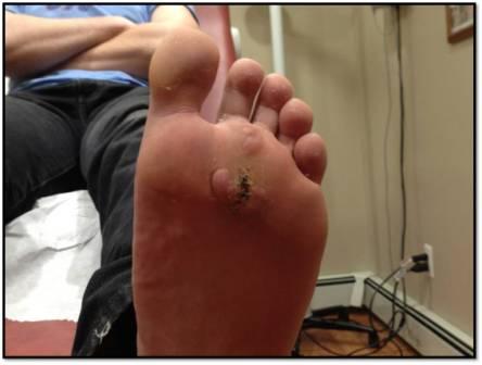
CHIEF COMPLAINT: Growth on the bottom of his left foot
CLINICAL HISTORY: 14 yo Caucasian male presented with growth on the bottom of his left foot for 3-4 months. He also endorses pain with ambulation. Previously, he had a series of curettages by podiatry, however the lesion continued to enlarge. Patient has a history of chronic macrocytosis and reticulocytopenia (bone marrow biopsy at age 10 revealed a non-clonal chromosome 15 deletion: 45 XY del(15)(q11.2)), developmental abnormalities, and Autism / Asperger’s disease.
PHYSICAL EXAM:
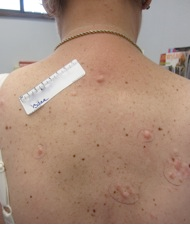
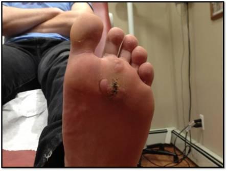
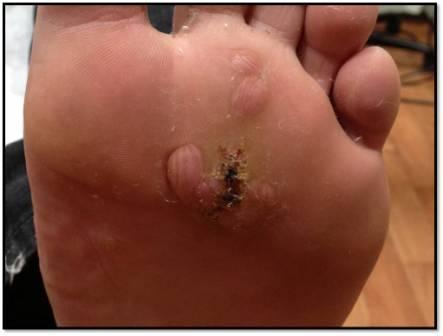
A full skin examination revealed one cafe au lait macule on the back. The plantar aspect of the left foot contained several flesh-colored cerebriform papules and nodules.
LABORATORY TESTS:
MRI brain: encephalomalacia and periventricular leukomalacia (localized areas of necrosis attributed to infarction or ischemia).
DERMATOHISTOPATHOLOGY:
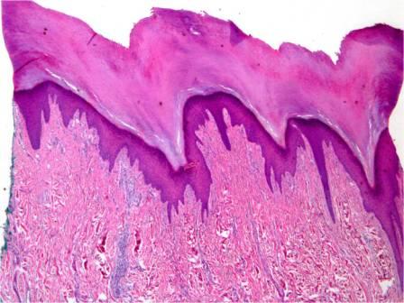
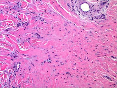
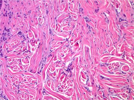
Histology revealed dense connective tissue beneath an acanthotic acantholytic epidermis. Stellate cells and entrapped adipose tissue were present in the dermis.
DIFFERENTIAL DIAGNOSIS:
1. Isolated plantar collagenoma
2. Plexiform neurofibroma (NF1)
3. SOLAMEN syndrome
4. Proteus syndrome
5. Multiple poromas

