CORRECT DIAGNOSIS:
Reed’s Syndrome
DISCUSSION:
Reed Syndrome (multiple cutaneous and uterine leiomyomatosis, MCUL) is an autosomal dominant condition characterized by the development of leiomyomata of the uterus in affected women, in addition to leiomyomata of the skin in both sexes. A subset of patients with Reed syndrome is at risk for renal cancer, in particular renal papillary type II cancer and renal collecting duct cancer. This disease variant is known as hereditary leiomyomatosis and renal cell cancer (HLRCC). To diagnose this condition, genetic testing for mutations of the fumarate hydratase (FH) gene must be performed.
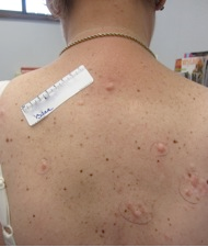
Clinically, cutaneous leiomyomatosis typically presents as painful nodules on the trunk and extremities during the second, third, or fourth decade of life in affected patients. Leiomyomas are characterized as smooth muscle tumors arising from arrectores pilorum muscles. The presence of these lesions is considered the most sensitive and specific clinical marker of HLRCC. In addition, female patients classically develop symptomatic leiomyomas of the uterus at a mean age of 20 to 35 years of age, often necessitating a hysterectomy to control symptoms. The risk of developing renal cancer in HLRCC is relatively low, estimated to be about 2-6%. However, it must be noted that when patients do develop renal carcinoma, it is usually metastatic by the time of diagnosis.
To diagnose this condition, genetic testing for mutations of the fumarate hydratase (FH) gene must be performed. The International Consortium identified fumarate hydratase as the gene responsible for MCUL/HLRCC in 2002. This gene is found on chromosome 1q 42-43 and encodes a Krebs cycle enzyme, which catalyzes the conversion of fumarate to malate. It has been hypothesized that the FH gene acts as a tumor suppressor gene; however, the exact mechanism by which tumorigenesis occurs is not known. While numerous defects in the FH gene have been identified, it has been proposed that frameshift mutations are responsible in the majority of patients with HLRCC.
TREATMENT:
Management of this condition involves symptomatic treatment of cutaneous lesions as well as close surveillance and monitoring for the development of renal tumors. Symptomatic treatment of leiomyomas includes, but is not limited to surgical excision, nifedipine, gabapentin, doxazosin, and botulinum toxin. Due to the aggressive nature of the renal cancer associated with this disease, initial screening with computed tomography or magnetic resonance imaging, in addition to periodic imaging, should be performed in patients who have tested positive for a FH mutation.
REFERENCES:
Mitchum, L. A., Tey, H. L., & Liao, W. (2012). A 46-year-old man with agminated papules on the buttock. Journal of the American Academy of Dermatology, 66(2), 337-339. https://doi.org/10.1016/j.jaad.2011.06.018
Alam, N. A., et al. (2005). Clinical features of multiple cutaneous and uterine leiomyomatosis: an underdiagnosed tumor syndrome. Archives of Dermatology, 141, 199-206. https://doi.org/10.1001/archderm.141.2.199
Alam, N. A., et al. (2005). Fumarate hydratase mutations and predisposition to cutaneous leiomyomas, uterine leiomyomas, and renal cancer. British Journal of Dermatology, 153, 11-17. https://doi.org/10.1111/j.1365-2133.2005.06453.x
Refae, A., et al. (2007). Hereditary leiomyomatosis and renal cell cancer: an unusual and aggressive form of hereditary renal carcinoma. Nature Clinical Practice Oncology, 4, 256-261. https://doi.org/10.1038/ncponc0785
Sanz-Ortega, J., et al. (2013). Morphologic and molecular characteristics of uterine leiomyomas in hereditary leiomyomatosis and renal cancer (HLRCC) syndrome. American Journal of Surgical Pathology, 37, 74-80. https://doi.org/10.1097/PAS.0b013e31826f250c


