CORRECT DIAGNOSIS:
Connective tissue disease [CTD] – Anti phospholipid Syndrome
DISCUSSION:
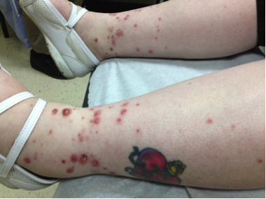
Most cases of cutaneous leukocytoclastic vasculitis (LCV) follow either an acute infection or exposure to a new medication1. Palpable purpura, as evidenced in this case, is a prime indicator of LCV and results from localized areas of hemorrhage occurring as the damaged vessels leak (red blood cells extravasation). These lesions are most often found on the ankles and lower legs with edema usually being an accompanying feature. They may coalesce into plaques with the possibility of ulceration. Other signs and symptoms that may be present include but are not limited to pruritus, fever, arthralgias, and arthritis. The disease is often self-limited with lesions usually resolving over the course of several weeks.
A thorough clinical history must be obtained focusing on recent medication/drug usage, recent infections, medical disease history, and overall review of systems. Accompanying laboratory testing should include a urinalysis, ASO titer, CBC, RF, ANA, Hepatitis panels, and if indicated, an ANCA, serum complements, cryoglobulins, and serum electrophoresis. These laboratory values along with clinical history and physical examination often can delineate whether the inciting issue follows a benign or more serious underlying condition. The investigation is directed at ruling out multisystem involvement with the most common systems being kidneys (50%), heart (50%), nervous system (40%) and GI tract (36%).
Biopsy taken in the first 24 hours may note DIF findings such as the presence of complement, immunoglobulins or fibrin deposition. The overall dysfunction of the small vessel usually is correlated with the circulating immune complexes. These lead to angiocentric inflammation of the post-capillary venule, expansion of the vessel wall, fibrin deposition with fibrinoid necrosis, and neutrophil infiltration1,2,3. Eosinophilia, if present, may correlate with medication being the offending agent.
Treatment for LCV often is directed at symptomatic improvement. Rest, elevation and analgesics are often the only required therapy. Oral antihistamines and corticosteroids may prove of useful benefit with the latter being indicated if any systemic signs or necrosis is present. Immunosuppressants including Methotrexate, Mycophenolate mofetil, Azathioprine, cyclosporine or cyclophosphamide may be required in particular patient scenarios3.
TREATMENT:
Prednisone 30mg PO BID with slow taper over one month. Additionally, Vistaril 25 PO qHS, and Jobst Stockings.
The lesions began to dissipate after two weeks of oral prednisone. The patient was lost to follow-up thereafter. Therefore, full resolution of the LCV after treatment completion is unknown. Furthermore, the possibility of recurrence remained as the inciting factor had yet to be determined. The patient denied symptoms of a recent infection. No signs correlating with her history of a “spider bite” were found on exam. Her only medication, Propranolol, had been prescribed for at least two years. However, onset of a vasculitic eruption after medication ingestion can be hours to years.5 Cases of LCV due to Beta-blockers have been reported. 6,7,8 Plans on follow-up included holding Propranolol and encouraging up-to-date age related cancer screening if lesions persisted or recurred.
REFERENCES:
Sunderkötter, C., Bonsmann, G., Sindrilaru, A., & Luger, T. (2005). Management of leukocytoclastic vasculitis. The Journal of Dermatological Treatment, 16(4), 193-206. https://doi.org/10.1080/09546630500112465.
PMID: 16117757
Gupta, S., Handa, S., Kanwar, A., Radotra, B., & Minz, R. (2009). Cutaneous vasculitides: Clinico-pathological correlation. Indian Journal of Dermatology, Venereology and Leprology, 75(4), 356-362. https://doi.org/10.4103/0378-6323.57863. PMID: 19714227
Tai, Y., Chong, A., Williams, R., Cumming, S., & Kelly, R. (2006). Retrospective analysis of adult patients with cutaneous leukocytoclastic vasculitis. The Australasian Journal of Dermatology, 47(2), 92-96. https://doi.org/10.1111/j.1440-0960.2006.00216.x. PMID: 16613676
Habif, T. P. (2010). Clinical dermatology. Elsevier.
James, W. D. (2011). Andrews’ Diseases of the skin. Saunders.
Wolf, R., Ophir, J., Elman, M., & Krakowski, A. (1989). Atenolol-induced cutaneous vasculitis. Cutis, 43(3), 231-233. PMID: 2465206
Pavlovic, M. D., Dragojevic Simic, V., Zolotarevski, L., Zecevic, R. D., & Vesic, S. (2007). Cutaneous vasculitis induced by carvedilol. Journal of the European Academy of Dermatology and Venereology, 21(7), 1004-1005. https://doi.org/10.1111/j.1468-3083.2007.02279.x. PMID: 17645706
Rustmann, W. C., Carpenter, M. T., Harmon, C., & Botti, C. F. (1998). Leukocytoclastic vasculitis associated with sotalol therapy. Journal of the American Academy of Dermatology, 38(1), 111-112. https://doi.org/10.1016/S0190-9622(98)70083-4. PMID: 9445047


