Presenter: Cathy Koger D.O., Steve Plumb D.O., Chris Cook D.O., Doug Richley D.O.
Dermatology Program: Northeast Regional Medical Center
Program Director: Lloyd Cleaver D.O.
Submitted on: January 4, 2014
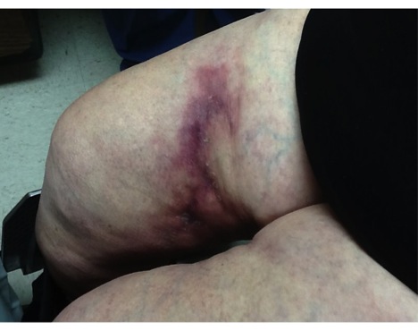
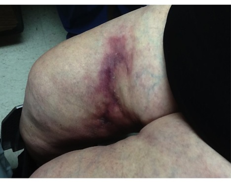
CHIEF COMPLAINT: rapidly expanding necrotic plaques of her lower extremities
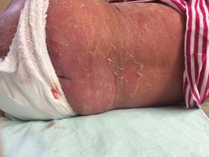
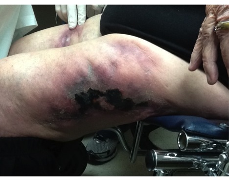
CLINICAL HISTORY: An 84-year-old Caucasian female presented with rapidly expanding necrotic plaques on her lower extremities that had developed over the past two months. The lesions, characterized by a reticulated pattern, were ulcerated, measuring approximately 15 cm, and extremely painful to the touch. They exhibited a firm, indurated texture and were accompanied by cord-like subcutaneous swellings. The patient had not received any previous treatment for these lesions. She resided in a local assisted living facility, where she received symptomatic care as needed. Her past medical history included hypercholesterolemia, for which she was prescribed Simvastatin, as well as a history of atrial fibrillation that required Warfarin. Notably, she had no prior history of skin cancer or other cutaneous issues.
PHYSICAL EXAM:
Scattered indurated, necrotic 15 cm ulcerated plaques with a reticulated configuration on the bilateral lower extremities. The lesions were extremely painful upon palpation. The patient had no cervical, axillary, or inguinal lymphadenopathy.
LABORATORY TESTS:
We ordered several lab tests at the initial visit. The labs were noteworthy for a normal BUN and creatnine, calcium, phosphorus, parathyroid, INR, and WBC count. Lastly, a deficiency in protein C was noted.
DERMATOHISTOPATHOLOGY:
An incisional biopsy revealed fibrin thrombi and the patient was admitted to the hospital for further workup. At the hospital, an excisional biopsy was performed on one of the ulcerated plaques which showed calcification within small and medium sized arterioles with intimal hyperplasia and fibrosis. In addition, there was a mixed inflammatory infiltrate as well as vascular microthrombi.
DIFFERENTIAL DIAGNOSIS:
1. Calciphylaxis
2. Cryoglobulinemia
3. DIC
4. Warfarin induced necrosis
5. Leukocytoclastic vasculitis

