CORRECT DIAGNOSIS:
Calciphylaxis
DISCUSSION:
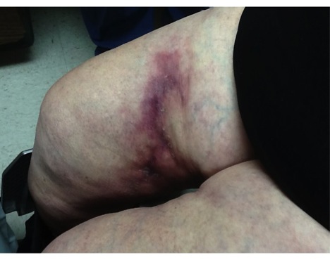
Calciphylaxis is a poorly understood and highly morbid disease that is characterized by progressive vascular calcification with ischemic necrosis of the skin and soft tissues. Early lesions feature violaceous reticulated patches that are subsequently followed by a grayish hue that signals tissue necrosis and ulcer formation. The lesions are characteristically extremely painful at any stage of development and proximal involvement has the worst prognosis. The overall mortality is estimated to be as high as 85% and is often secondary to sepsis and gangrene.
Patients with calciphylaxis are predominantly female and risk factors include end-stage renal disease, diabetes mellitus, obesity, and poor nutritional status. In addition, protein C dysfunction has been reported in calciphylaxis patients.
TREATMENT:
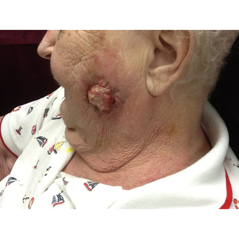
At the initial visit, two biopsies were performed, one for H&E and one for tissue C&S. The patient was admitted to our neighboring hospital for further workup and an array of labs were ordered. General surgery performed an excisional biopsy which was consistent with calciphylaxis. Furthermore, the patient was started on sodium thiosulfate by the hospitalist team and aggressive wound management was instituted. Interestingly, the patient was found to have colon adenocarcinoma as well as an acral lentiginous melanoma on her foot. She declined quickly while in the hospital and died within one month.
The current standard management of calciphylaxis includes the normalization of the calcium-phosphate product through the institutionalization of dialysis, phosphate binders, and parathyroidectomy if needed. Furthermore, meticulous wound care is important since a large proportion of patients die secondary to sepsis and gangrene.
Other proposed modalities include sodium thiosulfate, pamidronate, hyperbaric oxygen, and tissue plasminogen activator. Unfortunately, most of these recommendations are taken from small case reports and there is a lack of large scale evidence-based guidelines for practitioners to follow.
REFERENCES:
Bolognia, J. L., Jorizzo, J. L., & Rapini, R. P. (2012). Dermatology (3rd ed., pp. 731-732). Elsevier.
James, W. D., Berger, T. G., & Elston, D. M. (2011). Andrews’ Diseases of the Skin (11th ed., pp. 528, 819). Elsevier.
Eiser, A. R. (2013). Dialysis patients, atrial fibrillation, warfarin, and calciphylaxis: Outlier subsets and practice guidelines. American Journal of Medicine. https://doi.org/10.1016/j.amjmed.2013.10.020
PMID: 24388855
Ong, S., & Coulson, H. (2012). Diagnosis and treatment of calciphylaxis. Skinmed, 10(3), 166-170.
PMID: 22776784
Garcia, C. P., Roson, E., Peon, G., Abalde, M. T., & De La Torre, C. (2013). Calciphylaxis treated with sodium thiosulfate: Report of 2 cases. Dermatology Online Journal, 19(9), 19616.
PMID: 24078980


