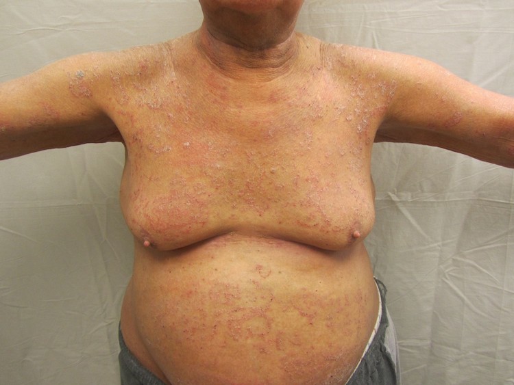
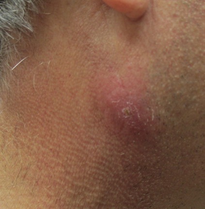
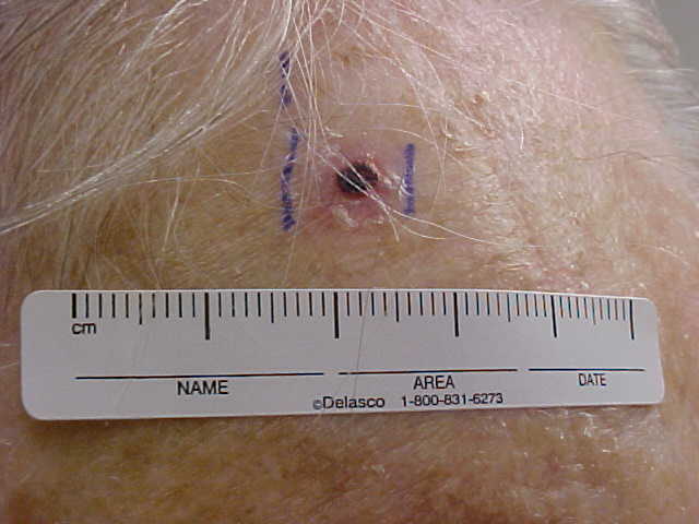
CORRECT DIAGNOSIS:
Pemphigus Foliaceous
DISCUSSION:
Pemphigus foliaceous (PF) is an acquired autoimmune blistering disease that is caused by IgG autoantibodies targeting keratinocyte desmosomes, specifically the desmosomal cadherin desmoglein-1 (Dsg-1). IgG4, the pathogenic subclass in most patients, inhibits the function of Dsg-1 resulting in the loss of cell-cell adhesion in keratinocytes and subsequent blister formation. Dsg-1 is expressed throughout the epidermis, but more intensely in the superficial layers. The split occurs in the superficial epidermis, mainly at the granular layer.
PF most commonly occurs between 40 and 60 years of age with no gender bias. It is characterized by flaccid bullae that very easily rupture to form erosions, crusts, and adherent scales. The lesions are often associated with pain, burning, or pruritic sensation. The Nikolsky sign is easily demonstrated. Distribution can be localized or widespread. The oral mucosa is very seldom, if ever, involved and patients generally do not appear ill.
The onset of PF has been associated with several drugs including penicillamine, captopril, enalapril, nifedipine, penicillin, and rifampin. It is thought that the implicated medications interact with the sulfhydryl groups in desmoglein. In the vast majority of patients, drug-induced PF will resolve after discontinuing the offending medication.
The primary histopathological finding is acantholysis in the upper epidermis, frequently within or adjacent to the granular layer. Intact blisters are rarely seen and only a few acantholytic cells at the roof or floor of the cleft may be present. Eosinophilic spongiosis can be seen in early lesions. However, histopathological findings alone are not enough to make a definitive diagnosis. The demonstration of IgG autoantibodies against the surface of keratinocytes is essential for diagnosis. Methods used for detection include direct immunofluorescence (DIF), indirect immunofluorescence (IIF) and immunoblotting and enzyme-linked immunosorbent assay (ELISA). DIF is the most reliable and sensitive diagnostic test. ELISA titers consistently correlate with disease activity and should be considered the best test for evaluating a patient’s response to therapy.
TREATMENT:
Randomized controlled trials for the treatment of PF are lacking. The literature does support oral corticosteroids as the backbone of therapy. Mycophenolate mofetil, azathioprine, and cyclophosphamide can be used as adjunctive therapy or as corticosteroid-sparing agents. Plasmapheresis and IVIG is reserved for refractory cases. Class I topical corticosteroids or calcineurin inhibitors may be effective for localized or mild disease. Medications should be tapered once clinical and serological remission is achieved.
This patient’s rash improved with a slow taper of prednisone beginning at 30mg and mycophenolate mofetil 500mg BID.
REFERENCES:
James, K. A., Culton, D. A., & Diaz, L. A. (2011). Diagnosis and clinical features of pemphigus foliaceus. Dermatologic Clinics, 29(3), 405-412. https://doi.org/10.1016/j.det.2011.03.001
Martin, L. K., Worth, V. P., Villaneuva, E. V., et al. (2011). A systematic review of randomized controlled trials for pemphigus vulgaris and pemphigus foliaceus. Journal of the American Academy of Dermatology, 64(5), 903-908. https://doi.org/10.1016/j.jaad.2010.07.041
Amagai, M., Komai, A., Hashimoto, T., et al. (1999). Usefulness of enzyme-linked immunosorbent assay using recombinant desmoglein 1 and 3 for serodiagnosis of pemphigus. British Journal of Dermatology, 140, 351-357. https://doi.org/10.1046/j.1365-2133.1999.02853.x


