CORRECT DIAGNOSIS:
Elephantiasis Nostras Verucosa
DISCUSSION:
Elephantiasis Nostras Verrucosa (ENV) is rare and represents end-stage development of two relatively more common conditions: edema and stasis. Prolonged edema and stasis are a common theme in many of the known risk factors namely: congestive heart failure, chronic venous insufficiency, obstructive tumors, previous surgery or trauma to lymphatic ducts, obesity, pulmonary hypertension, cellulitis and radiation(6) In patients with multiple risk factors like ours (heart failure, cellulitis, and pulmonary hypertension) there is a correlation with a more widespread disease (6). The further classification reveals almost all patients are morbidly obese (91%), have bilateral lower extremity involvement (86%), and concurrent cellulitis or other skin infections (86%).(6). ENV occurs alongside the skin changes of chronic venous insufficiency (stasis dermatitis, lipodermatosclerosis, ulceration) 71% of the time and has recently been recognized as an emerging risk factor for ENV(6). The classic ENV patient is most likely to be seen in cardiology or vascular surgery office(6). Those with mental illness are particularly vulnerable due to the inadvertent neglect of their health(7). However, ENV has been reported in nondependent areas such as the back, ears and abdomen making diagnosis challenging(8-11). Pathogenesis is due to the accumulation of interstitial protein-rich fluid. This inflammatory state and a localized weakened immune response. Cytokines from chronic inflammation stimulate fibroblasts and keratinocytes, transforming the initially soft pitting edema seen in stage 1 lymphedema, into hard fibrotic skin(12, 13). Histologic examination confirms this, showing fibrous tissue hyperplasia, pseudoepitheliomatous hyperplasia, dilated lymphatic spaces, and chronic inflammation(14). The weakened immune response provides a fertile ground and nidus for repeated bacterial infections further fibrosing the tissue(15). It appears that lypmhedema and stasis alone will cause indurated fibrotic skin but stop short of the cobblestoned and verrucous lesions characteristic of ENV . Bacterial superinfection is needed to develop these characteristic ENV lesions(14, 16).
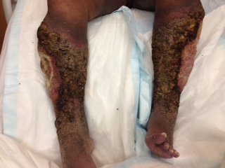
Unfortunately, we cannot further comment on whether a bacterial infection set off our patients ENV as he was a poor historian although, he was heavily colonized at the time of presentation. Chronic inflammation and infection imparts increased risk for malignancy development which needs to be ruled out by biopsy(17). Early disease is marked by fibrotic skin with clusters of indurated papillomatous nodules, which has prompted the use of the descriptive term- cobblestone. We feel our patient’s cobblestone-like nodules found above the knee, represented early disease, and was key in making the diagnosis (figure 2). These cobblestoned areas progress with verrucous projections and ulcerations in late-stage disease, as seen in our patient (Figure 1) .12 The differential diagnosis for ENV is venous stasis dermatitis, lipodermatosclerosis, pretibial myxedema, filariasis, lipedema, chromoblastomycosis, and papular mucinosis(15, 18). Pretyibial myxedmea can be ruled out by a TSH or lack of mucin in the biopsy specimen. Filarisis can be evaluated with a history of travel to endemic areas and peripheral blood smear or tissue stain identifying microorganisms. Lipedema, or the abnormal accumulation of fat, would show normal histology on biopsy and likely have a family history (15). Chromoblastomycosis would have culture positive for fungus and is more likely in tropical and subtropical climates. Venous stasis dermatitis can be ruled out clinically as it has pitting edema. Papular mucinosis would show acid glycosaminoglycan deposition on biopsy. Lipodermatosclerosis would have similar fibrotic skin but have fibrin deposits around capillaries.
TREATMENT:
Here we present one of the most extensive cases of ENV found in the literature. Common causes of lymphedema include heart failure, obstructive tumors, previous surgery or trauma to lymphatic ducts, obesity, pulmonary hypertension, cellulitis and radiation, all of which can provide the background for ENV to develop. The diagnosis continues to be clinical with testing focused at discovering the cause of lypmhedema. Treatment is focused on improving underlying lypmhedema. Adjunctive treatments include topical keratolytics, oral retinoids and surgical debridement in severe cases.
Treatment is not standardized but should be focused on relieving the lymphedema, this can be improved with extremity elevation and compression stockings or bandages(12, 13). Many different approaches, all with some degree of success, have been reported including topical keratolytics, oral retinoids(19, 20), complex lymphatic physical therapy(21), prophylactic antibiotics to prevent infections(10) and in extreme cases- surgical debridement(22, 23). Treatment for our patient consisted of compression stockings, leg elevation, and bleach baths – 1⁄2 cup bleach in a 40-gallon tub to prevent bacterial infection, antibiotic therapy was continued. A combination of Urea 40% cream BID and Clobetasol cream 0.05% BID was applied to verrucous areas to smooth the verrucous plaques. Aggressive long term compliance is needed to treat this disease.
REFERENCES:
1. Klaber R. Elephantiasis Nostras Verrucosa. Proc R Soc Med 1935;28:1171.
2. Castellani A. Elephantiasis Nostras (Non-filarial Elephantiasis): (Section of Tropical Diseases and Parasitology). Proc R Soc Med 1934;27:519-24.
3. Rockson SG. Lymphedema. Am J Med 2001;110:288-95.
4. Bolognia JL, Jorizzo JL , Rapini RP. Dermatology. Spain: Mosby Elsevier; 2008. p. 1602.
5. Lymphatic filariasis: the disease and its control. Fifth report of the WHO Expert Committee on
6. Dean SM, Zirwas MJ , Horst AV. Elephantiasis nostras verrucosa: an institutional analysis of 21 cases. J Am Acad Dermatol 2011;64:1104-10.
7. Simón Llanes J, Coll Vilar I, Tamarit Francés C , Niubó de Castro I. [Elephantiasis nostras verrucosa in a patient with major depressive disorder]. Semergen 2012;38:526-9. 8. Grant JM. Elephantiasis nostras verrucosa of the ears. Cutis 1982;29:441-4.
9. Berngard SC , Narayanan V. Elephantiasis nostras verrucosa of the pannus. J Gen Intern Med 2011;26:810.
10. Conley J , Anadkat M. What is your diagnosis? Elephantiasis nostras verrucosa of the back. Cutis 2013;91:63, 92.
11. Boyd J, Sloan S , Meffert J. Elephantiasis nostrum verrucosa of the abdomen: clinical results with tazarotene. J Drugs Dermatol 2004;3:446-8.
12. Fredman R , Tenenhaus M. Elephantiasis nostras verrucosa. Eplasty 2012;12:ic14.
13. Ryan TJ. Structure and function of lymphatics. J Invest Dermatol 1989;93:18S-24S.
14. Yang YS, Ahn JJ, Haw S, Shin MK , Haw CR. A case of elephantiasis nostras verrucosa. Ann Dermatol 2009;21:326-9.
15. Sisto K , Khachemoune A. Elephantiasis nostras verrucosa: a review. Am J Clin Dermatol 2008;9:141-
136 6.
16. Castellani A. Researches on Elephantiasis nostras and Elephantiasis tropica with special regard to their initial stage of recurring lymphangitis (Lymphangitis recurrens elephantogenica). J Trop Med Hyg 1969;72:89-97.
17. Ruocco E, Puca RV, Brunetti G, Schwartz RA , Ruocco V. Lymphedematous areas: privileged sites for tumors, infections, and immune disorders. Int J Dermatol 2007;46:662.
18. Liaw FY, Huang CF, Wu YC , Wu BY. Elephantiasis nostras verrucosa: swelling with verrucose appearance of lower limbs. Can Fam Physician 2012;58:e551-3.
19. Polat M , Sereflican B. A case of elephantiasis nostras verrucosa treated by acitretin. J Drugs Dermatol 2012;11:402-5.
20. Zouboulis CC, Biczó S, Gollnick H, Reupke HJ, Rinck G, Szabó M et al. Elephantiasis nostras verrucosa: beneficial effect of oral etretinate therapy. Br J Dermatol 1992;127:411-6.
21. Hinrichs CS, Gibbs JF, Driscoll D, Kepner JL, Wilkinson NW, Edge SB et al. The effectiveness of complete decongestive physiotherapy for the treatment of lymphedema following groin dissection for melanoma. J Surg Oncol 2004;85:187-92.
22. Ferrandiz L, Moreno-Ramirez D, Perez-Bernal AM , Camacho F. Elephantiasis nostras verrucosa treated with surgical débridement. Dermatol Surg 2005;31:731.
23. Iwao F, Sato-Matsumura KC, Sawamura D , Shimizu H. Elephantiasis nostras verrucosa successfully treated by surgical debridement. Dermatol Surg 2004;30:939-41.


