Presenter: Kristen Whitney DO, Michael Mortazie DO, Travis James DO, Carolyn Ellis DO
Dermatology Program: St Joseph Mercy Dermatology Program
Program Director: Daniel Stewart, DO
Submitted on: August 21, 2014
CHIEF COMPLAINT: 9 month history of a rapidly growing mass on the left maxilla.
CLINICAL HISTORY: A 70-year-old Caucasian male with an unremarkable past medical history presented with a nine-month history of a rapidly growing mass on the left maxilla. The patient denied any systemic symptoms including fever, chills, sweats, or rapid weight loss. He denied any facial pain, paresthesias, or bleeding to the area.
PHYSICAL EXAM:
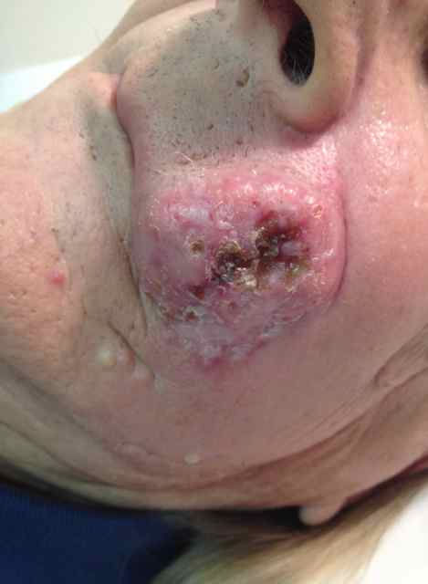
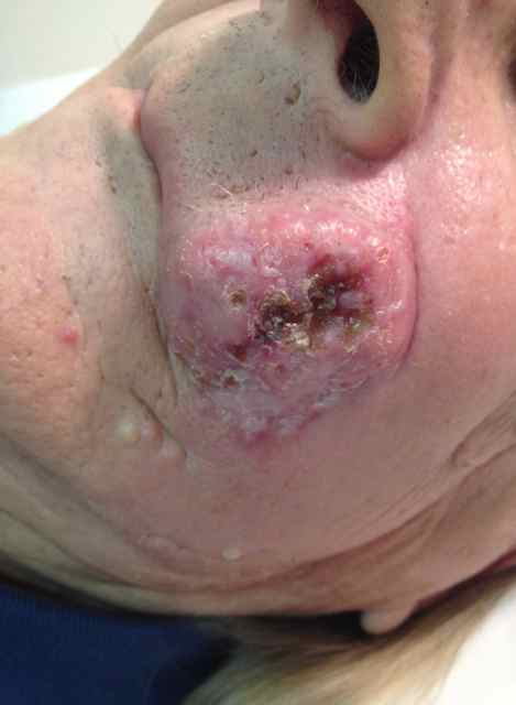
Physical examination of the left maxilla revealed a well-demarcated erythematous 4×4 cm tumor with a pearly border and central ulceration, extending from the superior border of the upper lip to the zygomatic arch.
LABORATORY TESTS:
PET imaging revealed increased activity in the left maxilla, with erosions into the alveolar ridge, left parotid gland, submandibular area, and left upper neck. A two-centimeter apical lung mass was found that was later proven to be non-malignant on CT guided biopsy.
DERMATOHISTOPATHOLOGY:
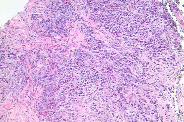
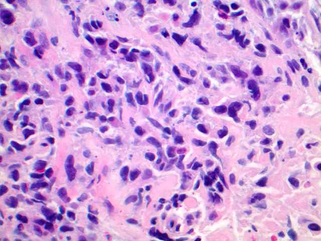
Three punch biopsies at the peripheral margin of the tumor demonstrated a poorly differentiated malignant neoplasm with large pleomorphic cells, enlarged hyperchromatic nuclei, and prominent nucleoli. Abundant mitoses and regions of necrosis were seen. In addition, there was a prominent eosinophilic infiltrate and epidermal hyperplasia. Tumor cells were CD3+, with a high concentration of CD4+ lymphocytes, along with a patchy infiltrate of CD8+ T-suppressor lymphocytes. Immunohistochemical stains were strongly positive for CD30; ALK was negative. In addition, cytokeratin AE1/3, S100, and MART-1 were all negative.
DIFFERENTIAL DIAGNOSIS:
1. Squamous Cell Carcinoma
2. Basal Cell Carcinoma
3. Deep Fungal Infection
4. Cutaneous Anaplastic Large-Cell Lymphoma
5. Cutaneous Metastasis

