CORRECT DIAGNOSIS:
Botryomycosis
DISCUSSION:
The term botryomycosis is derived from the Greek word botrys (meaning “bunch of grapes”) and mycosis (a misnomer, due to the presumed fungal etiology in early descriptions). Other terms used to describe botryomycosis include bacterial pseudomycosis, staphylococcal actinophytosis, granular bacteriosis, and actinobacillosis. The most frequent etiological agent is Staphylococcus aureus (40%), followed by Pseudomonas sp (20%). Other microorganisms reported include Escherichia coli, Proteus vulgaris, Bacillus spp, Actinobacillus lignieresii.
The pathogenesis of the disease has not been well established. It may be related to low virulence of agents, large local bacterial inoculum, change in specific cellular immunity (decreased number of T lymphocytes, like in agammaglobulinemia, aplastic anemia, agranulocytosis, and AIDS), or in humoral immune response (reduced IgA or increased IgE levels).
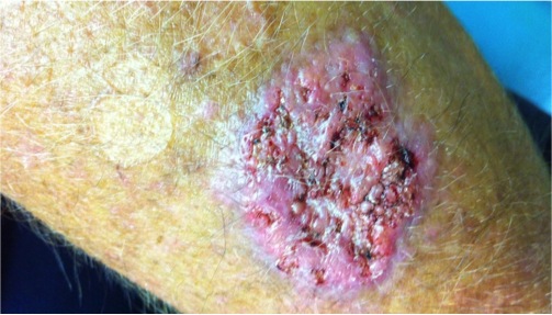
Similar to our case, most patients will present with a localized disease on the extremities that may be proceeded by trauma. Cutaneous botryomycosis is the most common form of botryomycosis and usually occurs following cutaneous inoculation of bacteria due to trauma, surgery, or in conjunction with the presence of a foreign body. Lesions characteristically develop slowly and may evolve and enlarge for several months and, rarely, even years.
The histopathologic appearance of botryomycosis is characterized by a central focus of necrosis surrounded by a chronic inflammatory reaction containing histiocytes, epithelioid cells, multinucleated giant cells, and fibrosis. Unlike the sulfur granules seen in actinomycosis (which contain filamentous branching organisms), the granules seen in botryomycosis contain bacteria surrounded by an eosinophilic matrix containing club-like projections. This histologic appearance is commonly referred to as the Splendore-Hoeppli phenomenon, although it may not always be present.
Diagnosing botryomycosis includes clinical suspicion and microbiologic studies. Isolation of the causative agent and susceptibility tests are essential to provide appropriate treatment
Although our patient’s biopsy lacked the classic Splendore-Hoeppli phenomenon, our final diagnosis remains botryomycosis. Strong consideration was also given to the possibility of this representing an unusual halogenoderma. Given the patient’s chronic exposure to Freon (halogenated hydrocarbons), this remained our working diagnosis pending the tissue culture results. However, with the heavy overgrowth of S. aureus represented histologically and via cultures and the patient’s response to a 2-week course of cephalexin 500mg QID cutaneous botryomycosis was favored.
TREATMENT:
In general, patients should receive antibiotic therapy until signs and symptoms of infection have resolved. Antibiotics for cutaneous disease include: oral trimethoprim-sulfamethoxazole (10-12 mg/kg /d), oral clindamycin (30-40mg/kg/d), cephalexin (500mg QID), Minocycline (100mg BID), doxycycline (100mg BID) or erythromycin (500 mg QID).
Our patient received and responded to a 2-week course of cephalexin 500mg QID.
REFERENCES:
Mehregan, D. A., Su, W. P., & Anhalt, J. P. (1991). Cutaneous botryomycosis. Journal of the American Academy of Dermatology, 24, 393–396. https://doi.org/10.1016/S0190-9622(91)70014-4
Bonifaz, A., & Carrasco, E. (1996). Botryomycosis. International Journal of Dermatology, 35, 381-388. https://doi.org/10.1111/j.1365-4362.1996.tb03494.x
Mechow, N., Göppner, D., Quist, S., et al. (2014). Cutaneous botryomycosis diagnosed long after an arm injury. Journal of the American Academy of Dermatology, 71(4), e155-156. https://doi.org/10.1016/j.jaad.2014.06.022
Schlossberg, D., Pandey, M., & Reddy, R. (1998). The Splendore-Hoeppli phenomenon in hepatic botryomycosis. Journal of Clinical Pathology, 51, 399. https://doi.org/10.1136/jcp.51.5.399
Askari, K., Seyed Saadat, S., Yousefi, N., Ghorbani, G., & Zargari, O. (2014). Cutaneous botryomycosis caused by Staphylococcus aureus in a patient with diabetes. International Journal of Dermatology, 53(4), 413-415. https://doi.org/10.1111/ijd.12123
Fernandes, N. C., Maceira, J. P., Knackfuss, I. G., & Fernandes, N. (2002). Botriomicose cutânea. Anais Brasileiros de Dermatologia, 77, 65-70. https://doi.org/10.1590/S0365-05962002000100011
James, W. D., Berger, T. G., et al. (2006). Andrews’ Diseases of the Skin: Clinical Dermatology. Saunders Elsevier.


