Presenter: Maren Gaul DO, Nathan Jackson DO
Dermatology Program: Western Reserve Hospital/Tri-County Dermatology
Program Director: Dr. Shield Wikas
Submitted on: November 2, 2014
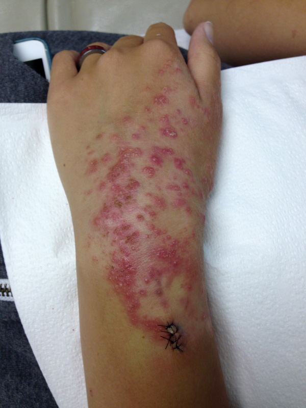
CHIEF COMPLAINT: Spreading rash from the left wrist (present for 11 months) to bilateral upper and lower extremities
CLINICAL HISTORY: 17-year-old Caucasian Female presented with complaint of spreading rash from the left wrist (present for 11 months) to bilateral upper and lower extremities in last week since biopsy of the wrist. The patient initially noted a rash on her left wrist in October 2013, which worsened on July 13, 2014. She admitted to pruritis and tenderness of the area of the left wrist with the involvement of the rash.
In July she went to minute clinic and was diagnosed with impetigo and given oral clindamycin for 2 days. She then went to urgent care, was diagnosed with poison ivy, and given prednisone, with some improvement. She also had received hydrocortisone valerate 0.2% and triamcinolone acetonide 0.5% as past therapy. Initial assessment at Tri-County Dermatology yielded a punch biopsy for H&E, which will be discussed below. She was also given halog 0.1% cream and a z-pak. Medical history and surgical history were negative. There were no recent infections, recent travel, or exposures to any new drugs or chemicals other than her previous treatments, and ROS such as arthritis, arthralgias, fever, or systemic involvement was negative.
PHYSICAL EXAM:
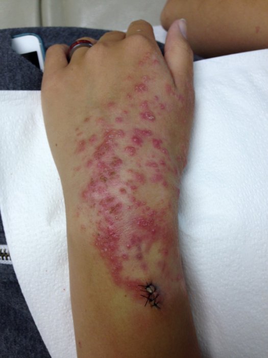
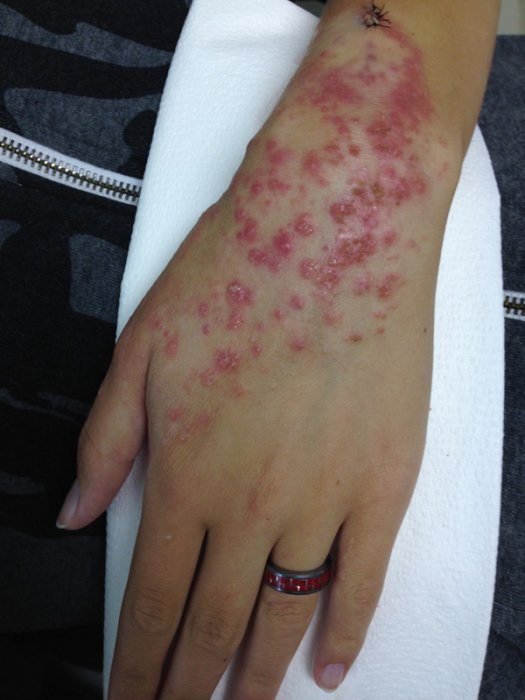
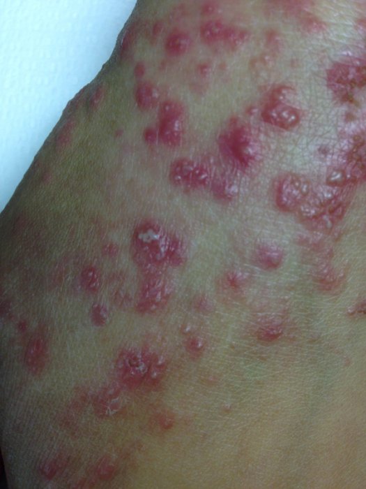
On July 29, 2014, a full skin exam revealed erythematous macules, papules, and pustules coalescing into patches and plaques on the dorsal and volar left wrist with the involvement of the proximal dorsal hand.
LABORATORY TESTS:
CBC and CMP were within normal limits
DERMATOHISTOPATHOLOGY:
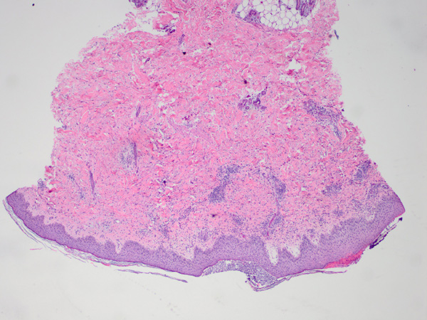
Punch biopsy of the left dorsal wrist with GMS demonstrated numerous fungal hyphae within subcorneal pustules of the stratum corneum. Sections had moderate irregular epidermal hyperplasia with moderate spongiosis. The granular cell layer was diminished with the overlying focal parakeratotic scale. There were multiple areas of subcorneal neutrophilic abscesses (pustules). Within the dermis was a moderate superficial perivascular lymphohistiocytic infiltrate with scattered neutrophils.
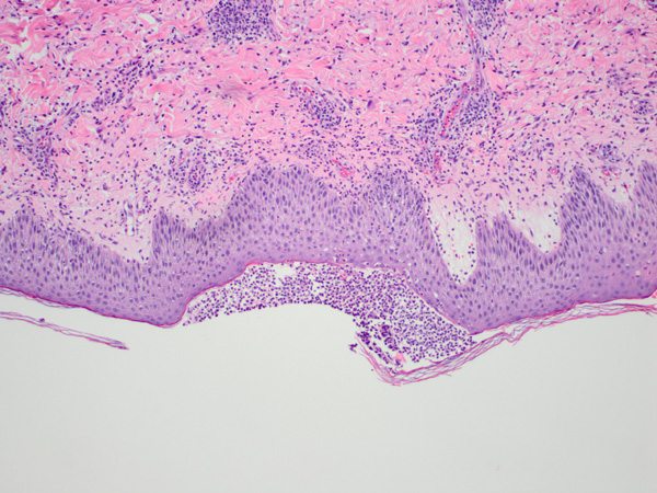
Detailed view of pustule:
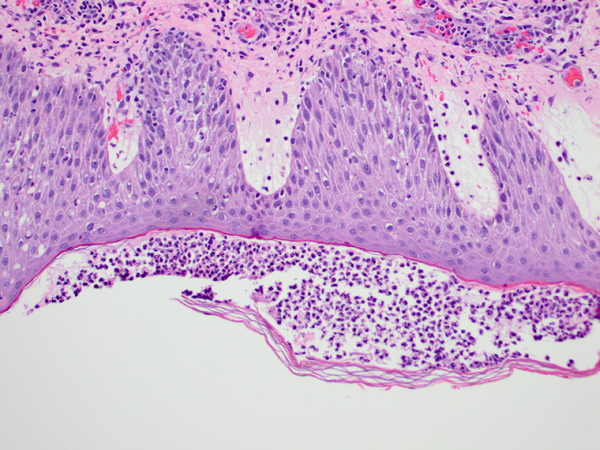
Subcorneal neutrophilic abscess, 400X:
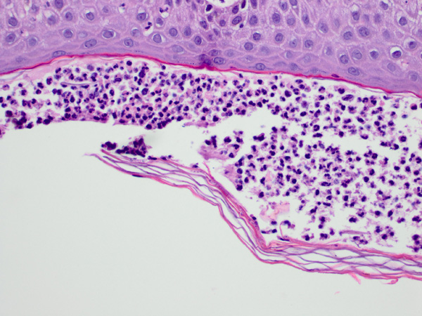
GMS demonstrated numerous fungal hyphae within subcorneal pustules of the stratum corneum:

DIFFERENTIAL DIAGNOSIS:
1. Pustular tinea
2. Sneddon-Wilkonson subcorneal pustular dermatosis
3. Tinea with secondary bacterial infection
4. Impetigo
5. Atopic dermatitis

