CORRECT DIAGNOSIS:
Scalp Arteriovenous Fistula with Intracranial Communication
DISCUSSION:
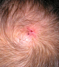
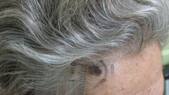
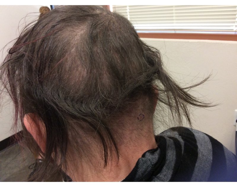
Scalp arteriovenous fistulas (S-AVFs) are characterized by abnormal connections between supplying arteries and draining veins in the subcutaneous plane of the scalp. The veins of a S-AVF undergo progressive aneurysmal dilatation as a result of abnormal hemodynamics. S-AVFs may present as a progressively enlarging pulsating mass on the scalp. Associated symptoms may include headache, local pain, bruits, tinnitus, and thrill. Hemorrhage and necrosis are less commonly associated.
S-AVFs may occur with or without intracranial communication. Spontaneous S-AVFs with intracranial communication are uncommon and their etiology is unclear. Spontaneous S-AVFs may form as congenital malformations or may be idiopathic. Most patients remain asymptomatic until puberty and typically seek medical attention after the second decade. Factors increasing the circulation through the S-AVF such as trauma, pregnancy, hormonal changes, endocrine stimulation, and inflammation prompt the development of symptoms.
S-AVFs may also be caused by trauma. S-AVFs without intracranial communication have been reported following hair transplantation. S-AVFs with intracranial communication may develop months to years after skull fracture or craniotomy. True spontaneous S-AVFs are difficult to differentiate from traumatic S-AVFs other than by history alone.
There are three proposed hypotheses for the development of spontaneous S-AVFs. The first is the persistence of primitive arteriovenous communication and capillary agenesis. The second suggests that S-AVFs originate from vascular hamartomas. The third is the formation of a fistula at the site of arteriovenous crossing.
The diagnosis of a S-AVF is confirmed with imaging studies. A Doppler ultrasound will help to initially detect that a lesion is vascular in nature. Intra-arterial digital subtraction angiography is the gold standard imaging technique and is necessary to delineate the feeding arteries and the draining channels, as well as possible communication with intracranial vasculature.
There is controversy regarding the appropriate treatment of S-AVFs. The prognosis for S-AVFs is extremely variable and the decision to treat is based upon the patient’s symptoms and risk of exsanguinating hemorrhage. Neurosurgical approaches to the management of S-AVFs include ligation of the feeding arteries, surgical resection, electrothrombosis, and embolization. Endovascular intervention is increasingly being used as a primary treatment or as a preoperative adjunct to surgery. In cases with intracranial communication, the intracranial component is treated first.
We report this case to emphasize the importance of keeping S-AVFs on the dermatologic differential diagnosis of a scalp mass. We recommend taking a thorough history, inquiring about the associated S-AVF symptoms, palpating suspicious scalp masses for bruits, and appropriate imaging if indicated. S-AVFs should be referred to neurosurgery or interventional neuroradiology for evaluation and possible treatment.
TREATMENT:
The patient was treated by interventional neuroradiology with intravascular embolization, which resulted in complete resolution of the scalp nodule.
REFERENCES:
Bernstein, J., Podnos, S., & Leavitt, M. (2011). Arteriovenous fistula following hair transplantation. Dermatologic Surgery, 37(6), 873-875. https://doi.org/10.1111/j.1524-4725.2011.01965.x
Kumar, R., Sharma, G., & Sharma, B. S. (2012). Management of scalp arteriovenous malformation: Case series and review of literature. British Journal of Neurosurgery, 26(3), 371-377. https://doi.org/10.3109/02688697.2012.670154
Senoglu, M., Yasim, A., Gokce, M., & Senoglu, N. (2008). Nontraumatic scalp arteriovenous fistula in an adult: Technical report on an illustrative case. Surgical Neurology, 70(3), 194-197. https://doi.org/10.1016/j.surneu.2007.07.012


