CORRECT DIAGNOSIS:
Fixed Drug Eruption
DISCUSSION:
After the biopsy results returned, a careful review of the patient’s medical history revealed that each episode was produced by the same event –recreational use of marijuana.
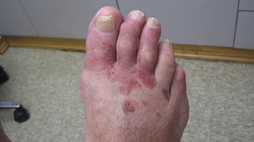
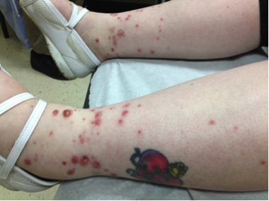
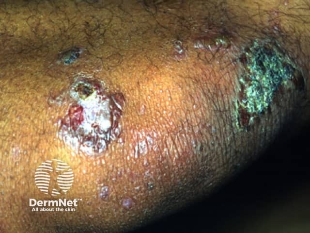
Drug eruptions are one of the most common cutaneous disorders encountered by dermatologists, representing 2 to 3% of all dermatological issues. FDE is a form of drug allergy that presents as single, or multiple rounds, sharply demarcated dusky red lesions several centimeters in diameter that occur at the same sites after each administration of the inciting drug. Pruritis and burning are often associated with symptoms. The average age of onset is approximately 30 years old and the most commonly implicated medication is trimethoprim-sulfamethoxazole. Between the time when the individual is first exposed to the medication and development of the first lesion, a variable refractory period can exist for a week, months, or even years. With subsequent exposure, lesions appear within thirty minutes to eight hours. Most commonly, the lesions heal with residual hyperpigmentation. Our patient, presented with the classic pigmented FDE.
While generally only a solitary lesion appears on first exposure, repeated administration of the medication can lead to new lesions or an increase in the size of the original lesions. Although they can occur anywhere on the skin, FDE’s most commonly occur on the glans penis, lips, palms, soles, and groin area. Overall, the legs are most commonly affected in women and the genitalia are most commonly affected in men.
Histological examination displays two possible scenarios depending on when the biopsy was done. In lesions that are only one to two days old, hydropic degeneration of basal keratinocytes with dyskeratotic cells in the epidermis and exocytosis of mononuclear cells are seen. Healed hyperpigmented lesions often demonstrate pigmentary incontinence revealing dermal melanophages with little perivascular infiltration of inflammatory cells. To identify the culprit of the FDE, provocation tests can be done with the patch test being the most commonly used method as long as it is placed over a previously involved site and the patient is not in the refractory period. Challenging a patient with an oral provocation test has been associated with generalized bullous lesions in some cases. In our case, we did not re-challenge the patient with the suspected drug due to legal concerns.
TREATMENT:
Treatment consists of cessation of suspected drugs with the use of topical steroids and systemic antihistamines. Extensive lesions or those with bullae may require systemic corticosteroids. Post-inflammatory hyperpigmentation can be treated with hydroquinone bleaching creams. In our patient, a short course of class 5 topical corticosteroid therapy resulted in complete resolution of the lesions, and patient was advised to abstain from marijuana.
REFERENCES:
Pai, V. V., Bhandari, P., Kikkeri, N. N., Athanikar, S. B., & Sori, T. (2012). Fixed drug eruption to fluconazole: A case report and mini-review of literature. Indian Journal of Pharmacology, 44(5), 643-645. https://doi.org/10.4103/0253-7613.100792 [PMID: 23248378]
Brocq, L. (1894). Eruption erythemato-pigmentee fixe due a l’antipyrine. Annales de Dermatologie et de Vénéréologie, 5, 308–313.
Lee, A. Y. (2000). Fixed drug eruptions: Incidence, recognition and avoidance. American Journal of Clinical Dermatology, 1(5), 277-285. https://doi.org/10.2165/00128071-200001050-00003 [PMID: 15050981]
Stritzler, C., & Kopf, A. W. (1960). Fixed drug eruption caused by 8-chlorotheophylline in Dramamine with clinical and histologic studies. Journal of Investigative Dermatology, 34, 319-330. https://doi.org/10.1038/jid.1960.58
Gendernalik, S. B., & Galeckas, K. J. (2009). Fixed drug eruptions: A case report and review of literature. Cutis, 84(4), 215-219.
Ozkaya-Bayazit, E., Bayazit, H., & Ozarmagan, G. (2000). Drug-related clinical pattern in fixed drug eruption. European Journal of Dermatology, 10(4), 288-291. [PMID: 10947085]
Liu, S. W., Lien, M. H., & Fenske, N. A. (2010). The effects of alcohol and drug abuse on the skin. Clinical Dermatology, 28(4), 391-399. https://doi.org/10.1016/j.clindermatol.2010.01.002 [PMID: 20472268]
Substance Abuse and Mental Health Services Administration, Center for Behavioral Health Statistics and Quality. (2014). The NSDUH Report: Substance Use and Mental Health Estimates from the 2013 National Survey on Drug Use and Health: Overview of Findings. SAMHSA: Rockville, MD.


