Presenter: Natalie Edgar DO, Dawnielle Endly DO, Joseph Dyer DO
Dermatology Program: Largo Medical Center / NSUCOM
Program Director: Richard Miller DO
Submitted on: May 1, 2015
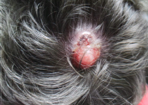
CHIEF COMPLAINT: Scalp nodule enlarging over 5 weeks
CLINICAL HISTORY: A 49-year-old Caucasian male presented with a scalp nodule enlarging over 5 weeks. The nodule was intermittently bleeding but non-tender to palpation. No previous treatment. Past medical history was pertinent for cystic fibrosis necessitating bilateral lung transplants in 2009. Current medications included mycophenolate mofetil 1.5 g twice daily, tacrolimus 1 mg twice daily, and prednisolone 5 mg daily. He had no history of visceral malignancy.
PHYSICAL EXAM:
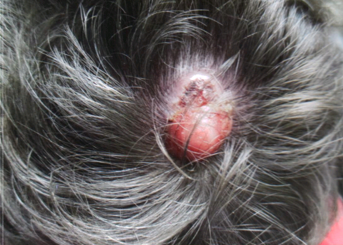
Clinical examination revealed a 2.7 cm ulcerated, exophytic, pink, keratotic nodule on the posterior scalp. No cervical or supraclavicular lymphadenopathy was appreciable.
LABORATORY TESTS: N/A
DERMATOHISTOPATHOLOGY:
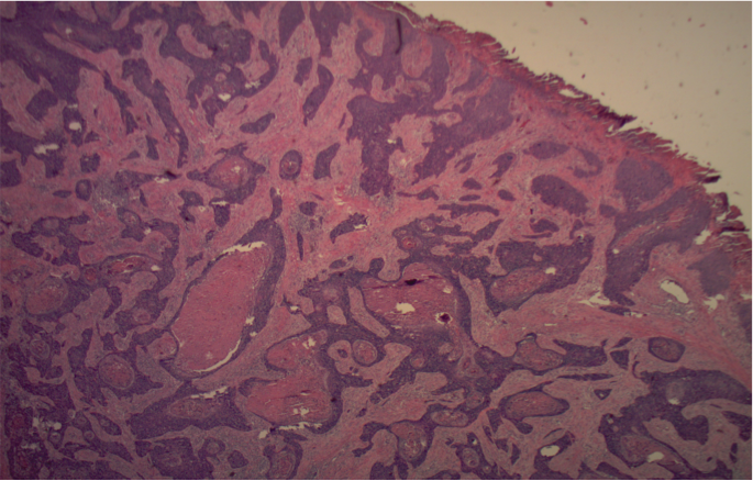
Low-power microscopy showed a proliferation of atypical epithelium emanating from the intraepidermal portion of the eccrine sweat glands and extending into the dermis. The high-power review demonstrated keratinizing epithelioid cells with intercellular bridges. No areas of lymphovascular or perineurial invasion were noted. There were < 14 mitotic figures per high-power field. Tumor depth was noted to be at least 6.5 mm.
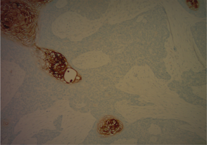
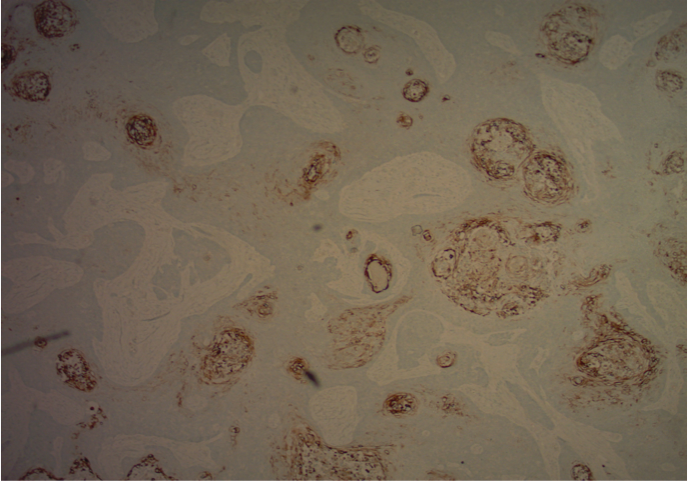
After special stains, cell populations were positive for CEA and CK7, confirming eccrine duct differentiation.
DIFFERENTIAL DIAGNOSIS:
1. Pyogenic granuloma
2. Basal cell carcinoma
3. Squamous cell carcinoma
4. Amelanotic melanoma
5. Cutaneous metastases

