CORRECT DIAGNOSIS:
TUMID LUPUS ERYTHEMATOSUS (TLE)
DISCUSSION:
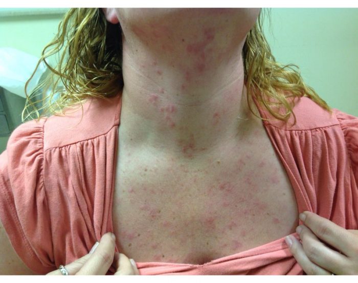
Tumid lupus erythematosis(TLE) is an uncommon variant of chronic cutaneous lupus. It is typically more common in young females and presents in a photo distributed pattern on the face, neck, chest, back, and extensor surfaces on the upper arms. The lesions commonly appear as asymptomatic erythematous, non-scaly papules, plaques, and nodules. Most patients have a negative serologic workup, as in our patient’s lab work above, including negative ANA, Anti-Ro and Anti-La. Common histopathological findings include increased dermal mucin, superficial and deep perivascular and periadnexal lymphocytic infiltrates. Interface changes are uncommon in tumid lupus erythematosus. In contrast, systemic lupus and discoid lupus have the involvement of the dermal-epidermal junction with vacuolar degeneration and necrotic keratinocytes.
TREATMENT:
In general, TLE is not associated with any systemic diseases, the lesions are self-limited and have a good prognosis. Treatment typically includes the anti-malarial therapy hydroxychloroquine. Our patient was placed on hydroxychloroquine 200mg twice daily. Her clinical improvement is pending follow up. Smoking cessation was advised as this may worsen cutaneous lupus. Sun protective measures were also stressed to our patient.
REFERENCES:
Maize, J.C., & Costner, M. (2010). Tumid lupus erythematosus: A form of lupus erythematosus. Archives of Dermatology, 146(4), 451; author reply 450-1.
Callen, J.P. (2009). Clinically relevant information about cutaneous lupus erythematosus. Archives of Dermatology, 145(3), 316-319.
Perez, G., Kowalczyk, J.P., Montie, D., & Nousari, C.H. (2009). Tumid lupus erythematosus at the site of a scar. Archives of Dermatology, 145(3), 343-344.
Pacheco, T.R., Spates, S.T., & Lee, L.A. (2002). Unilateral tumid lupus erythematosus. Lupus, 11(6), 388-391.


