Presenter: Jessica Vincent, DO
Dermatology Program: OhioHealth O’bleness Hospital
Program Director: Dawn Sammons, DO, FAOCD
Submitted on: April 13, 2016
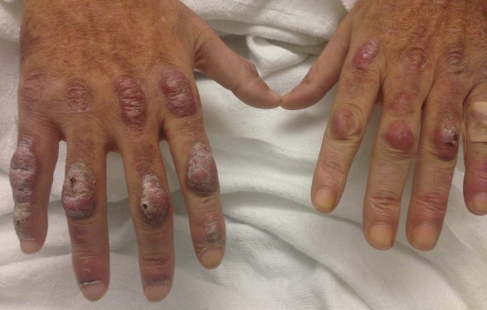
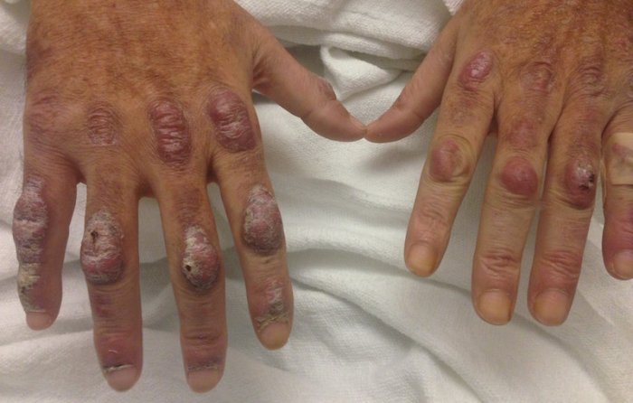
CHIEF COMPLAINT: Burning and stinging red nodules on the dorsum of his hands x 1 year
CLINICAL HISTORY: A 57-year-old male presented to the current authors complaining of burning and stinging red nodules on the dorsum of his hands for about 1 year. He also admitted to the persistence of an episodic rash over the lower legs and bilateral flanks he had originally presented with 7 years prior. He was briefly treated with an oral prednisone taper and topical corticosteroids including triamcinolone 0.1% cream and clobetasol 0.05% cream without improvement. A biopsy 7 years prior revealed leukocytoclastic vasculitis (LCV) with prominent eosinophils. At the time, it was felt his skin findings were a manifestation of drug hypersensitivity, likely to opioid use. The patient was subsequently lost to follow up.
PHYSICAL EXAM:
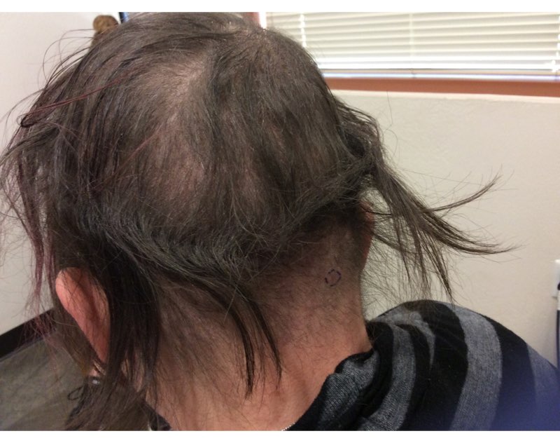
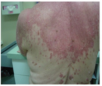
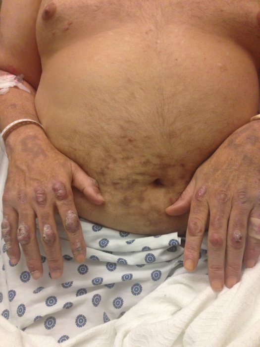
On exam were deep red to violaceous discrete nodules and plaques with overlying hyperkeratosis involving all distal and proximal interphalangeal joints of the hands and extensor elbows. On the bilateral posterior arms, anterior legs and periumbilical area were deeply erythematous papules and plaques with background hyperpigmentation. Across his low back and bilateral flanks were erythematous papules with central hemorrhagic crusting.
LABORATORY TESTS:
Pertinent laboratory findings included a positive hepatitis B surface antigen with hepatitis B DNA value 4313876 IU/mL and a HBV quantitative PCR value of 6.64 units (reference range <1
DERMATOHISTOPATHOLOGY:
Three 4-mm punch biopsies were performed from the left 5th digit, left posterior arm, and left flank. The tissue of the left 5th digit showed an intradermal vascular proliferation with a concentric, “onion skin” pattern in a background of increased fibrosis. The blood vessels showed focal fibrinoid necrosis. The biopsy of the left posterior arm showed an intradermal vascular proliferation with an associated mild acute and chronic perivascular inflammation. The left flank biopsy showed leukocytoclastic vasculitis with focal epidermal necrosis.
DIFFERENTIAL DIAGNOSIS:
1. Henoch-Schonlein purpura
2. Cryoglobulinemia
3. Sarcoidosis
4. Multicentric reticulohistiocytosis
5. Livedoid vasculopathy

