CORRECT DIAGNOSIS:
Erythema elevatum diutinum
DISCUSSION:
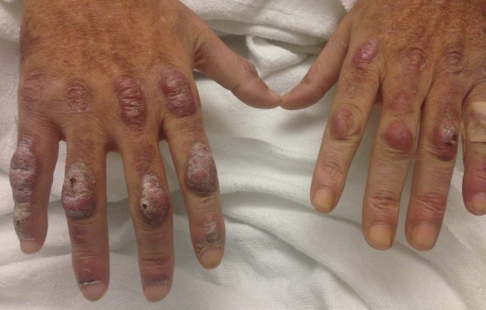
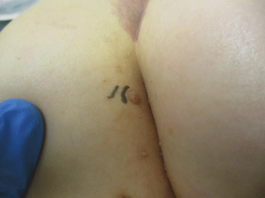
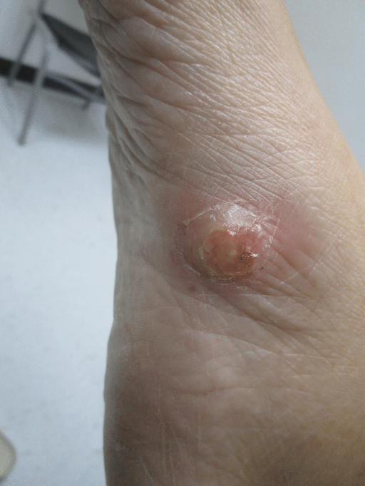
Erythema elevatum diutinum represents a rare form of chronic cutaneous small vessel vasculitis. Originally described by Hutchinson and Bury as symmetric, purpuric nodules of the skin,7,8 it was later named by Radcliffe-Crocker and Williams in 1894.9 The disease classically presents as firm, fixed red-brown to violaceous papules, plaques and nodules affecting the extensor extremities.1 Lesions are most commonly found symmetrically overlying joints of the hands, feet, elbows and knees as well as the Achilles tendon and buttocks. 3 Less common locations have been described including palms and soles, face, 10, 11 trunk 12 and periauricular region.1 While typically asymptomatic, sensations such as burning, stinging and pruritus have been noted.1 Our patient was unique as in addition to typical lesions of EED, he presented with crusted papules on the flanks and violaceous papules of the lower legs and periumbilicus.
Originally associated with Streptococcus as isolated from EED lesions,13, 3 additional infectious etiologies include viral hepatitis,4,5,6 HHV-6 14 and HIV.1, 15 Hepatitis B and C are well-known to be associated with EED, however, only previously reported in patients with concomitant HIV infection. EED has also been described in relation to myeloproliferative disorders and hematologic malignancies such as IgA myeloma,16 non-Hodgkin lymphoma,17 chronic lymphocytic leukemia 18 and hypergammaglobulinemia.19 In a study of 13 patients with EED by Yiannais et al, 4 had associated underlying IgA monoclonal gammopathy.2 Autoimmune conditions such as rheumatoid arthritis20, ulcerative colitis,21 relapsing polychondritis 22 and systemic lupus erythematosus 23 have also been implicated.
While the precise pathogenesis of EED remains unknown, it has been suggested that a complement cascade initiated by immune-complex deposition in post-capillary venules induces a leukocytoclastic vasculitis.24,25 Chronic antigenic exposure or high antibody levels26 in the face of infections, autoimmune disease, or malignancy may incite this immune-complex reaction. Skin lesions seen in association with hepatitis reflect circulating immune-complex deposition in vessel walls causing destruction. It has been postulated that the duration of immune complexemia may be sufficient to account for the differences in the type of vascular injury seen in acute versus chronic infection.27
EED may present on a histopathologic spectrum of LCV, as manifested in our patient. Early lesions show predominantly polymorphonuclear cells with nuclear dust pattern in a wedge-shaped infiltrate with fibrin deposition in the superficial and mid-dermis.2,3 Later lesions show vasculitis in addition to dermal aggregates of lymphocytes, neutrophils, fibrosis and areas of granulation tissue. The fibrosis may be dense and comprised of fibroblasts and myofibroblasts.28 Newly formed vessels within the granulation tissue have been postulated to be more susceptible to immune-complex deposition, thus potentiating the process.1,29
TREATMENT:
Spontaneous resolution of EED may occur, albeit after a prolonged and recurrent course of up to 5-10 years.30 Treatment of the underlying cause, when identified, remains paramount. First-line therapy includes dapsone, shown to be effective in reducing lesion size to complete resolution in 80% of the 47 cases in the literature reviewed by Momen and colleagues.31 Dapsone monotherapy tends to be less effective in treating nodular lesions associated with HIV-positivity, likely due to the extensive fibrosis.4,31 Combination therapy with dapsone and a sulfonamide,32 niacinaminde and tetracycline,33 colchicine,34 or surgical excision35 may be necessary in more resistant cases.
REFERENCES:
Gibson, L. E., & el-Azhary, R. A. (2000). Erythema elevatum diutinum. Clinics in Dermatology, 18(3), 295-299.
Yiannias, J. A., el-Azhary, R. A., & Gibson, L. E. (1992). Erythema elevatum diutinum: A clinical and histopathologic study of 13 patients. Journal of the American Academy of Dermatology, 26(1), 38-44.
Wilkinson, S. M., English, J. S., Smith, N. P., Wilson-Jones, E., & Winkelmann, R. K. (1992). Erythema elevatum diutinum: A clinicopathological study. Clinical and Experimental Dermatology, 17(2), 87-93.
Fakheri, A., Gupta, S. M., White, S. M., Don, P. C., & Weinberg, J. M. (2001). Erythema elevatum diutinum in a patient with human immunodeficiency virus. Cutis, 68, 41-42.
Kim, H. (2003). Erythema elevatum diutinum in an HIV-positive patient. Journal of Drugs in Dermatology, 2(4), 411-412.
Revenga, F., Vera, A., Muñoz, A., De la Llana, F. G., & Alejo, M. (1997). Erythema elevatum diutinum and AIDS: Are they related? Clinical and Experimental Dermatology, 22, 250-251.
Hutchinson, J. (1888). On two remarkable cases of symmetrical purple congestion of the skin in patches, with induration. British Journal of Dermatology, 1, 10-15.
Bury, J. S. (1889). A case of erythema with remarkable nodular thickening and induration of the skin associated with intermittent albuminuria. Illustrated Medical News, 3, 145-149.
Crocker, H. R., & Williams, C. (1894). Erythema elevatum diutinum. British Journal of Dermatology, 6, 33-38.
Barzegar, M., Davatchi, C. C., Akhyani, M., Nikoo, A., Daneshpazhooh, M., & Farsinejad, K. (2009). An atypical presentation of erythema elevatum diutinum involving palms and soles. International Journal of Dermatology, 48, 73-75.
Futei, Y., & Konohana, I. (2000). A case of erythema elevatum diutinum associated with B-cell lymphoma: A rare distribution involving palms, soles, and nails. British Journal of Dermatology, 142, 116-119.
Ben-Zvi, G. T., Bardsley, V., & Burrows, N. P. (2014). An atypical distribution of erythema elevatum diutinum. Clinical and Experimental Dermatology, 39, 269-270.
Weidman, F. D., & Besancon, J. H. (1929). Erythema elevatum diutinum. Role of streptococci, and relationship to other rheumatic dermatoses. Archives of Dermatology and Syphilology, 20, 593-620.
Drago, F., Semino, M., Rampini, P., Lugani, C., & Rebora, A. (1999). Erythema elevatum diutinum in a patient with human herpesvirus 6 infection. Acta Dermato-Venereologica, 79(1), 91-92.
Muratori, S., Carrera, C., Gorani, A., & Alessi, E. (1999). Erythema elevatum diutinum and HIV infection: A report of five cases. British Journal of Dermatology, 141(2), 335-338.
Archimandritis, A. J., Fertakis, A., Alegakis, G., Bartsokas, A., & Melissinos, K. (1977). Erythema elevatum diutinum and IgA myeloma: An interesting association. British Medical Journal, 2, 613-614.
Hatzitolios, A., Tzellos, T. G., Savopoulos, C., Tzalokostas, V., Psomas, E., Apostolopoulou, M., & Papadopoulos, A. (2008). Erythema elevatum diutinum with rare distribution as a first clinical sign of non-Hodgkin’s lymphoma: A novel association? Journal of Dermatology, 35, 297-300.
Delaporte, E., Alfandari, S., Fenaux, P., Piette, F., & Bergoend, H. (1994). Erythema elevatum diutinum and chronic lymphocytic leukaemia. Clinical and Experimental Dermatology, 19, 188-189.
Miyagawa, S., Kitamura, W., Morita, K., Saishin, M., & Shirai, T. (1993). Association of hyperimmunoglobulinaemia D syndrome with erythema elevatum diutinum. British Journal of Dermatology, 128, 572-574.
Collier, P. M., Neill, S. M., Branfoot, A. C., & Staughton, V. (1990). Erythema elevatum diutinum – A solitary lesion in a patient with rheumatoid arthritis. Clinical and Experimental Dermatology, 15, 394-395.
Buahene, K., Hudson, M., Mowat, A., Smart, L., & Ormerod, A. D. (1991). Erythema elevatum diutinum—An unusual association with ulcerative colitis. Clinical and Experimental Dermatology, 16, 204-206.
Bernard, P., Bedane, C., Delrous, J. L., Catanzano, G., & Bonnetblanc, J. M. (1992). Erythema elevatum diutinum in a patient with relapsing polychondritis. Journal of the American Academy of Dermatology, 26, 312-315.
Hancox, J. G., Wallace, C. A., Sangueza, O. P., & Graham, G. F. (2004). Erythema elevatum diutinum associated with lupus panniculitis in a patient with discoid lesions of chronic cutaneous lupus erythematosus. Journal of the American Academy of Dermatology, 50, 652-653.
Haber, H. (1955). Erythema elevatum diutinum. British Journal of Dermatology, 67, 121-145.
Katz, S. I., Gallin, J. L., Hertz, K. C., Fauci, A. S., & Lawley, T. J. (1977). Erythema elevatum diutinum: Skin and systemic manifestations, immunologic studies, and successful treatment with dapsone. Medicine, 56, 443-455.
Walker, K. D., & Badame, A. J. (1990). Erythema elevatum diutinum in a patient with Crohn’s disease. Journal of the American Academy of Dermatology, 22, 948-952.
Popp, J. W., Harrist, T., Dienstag, J. L., Bhan, A. K., Wands, J. R., LaMont, J. T., Mihm, M. C. (1981). Cutaneous vasculitis associated with acute and chronic hepatitis. Archives of Internal Medicine, 141, 623-629.
Lee, A. Y., Nakagawa, H., Nogita, T., & Ishibashi, Y. (1989). Erythema elevatum diutinum: An ultrastructural case study. Journal of Cutaneous Pathology, 16(4), 211-217.
LeBoit, P. E., Yen, T. S., & Wintroub, B. (1986). The evolution of lesions in erythema elevatum diutinum. American Journal of Dermatopathology, 8(5), 392-402.
Soubeiran, E., Wacker, J., Hausser, I., & Hartschuh, W. (2008). Erythema elevatum diutinum with unusual clinical appearance. Journal of the German Society of Dermatology, 6(4), 303-305.
Momen, S. E., Jorizzo, J., & Al-Niaimi, F. (2014). Erythema elevatum diutinum: A review of presentation and treatment. Journal of the European Academy of Dermatology and Venereology, 28(12), 1594-1602.
Vollum, D. I. (1968). Erythema


