Presenter: Duggan C., Jajou P.
Dermatology Program: Beaumont Hospital Trenton
Program Director: Steven Grekin DO
Submitted on: April 29, 2016
CHIEF COMPLAINT: Pruritic spreading rash
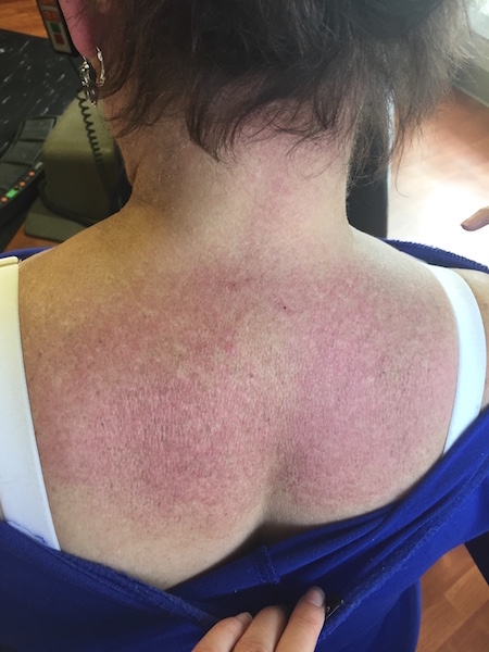
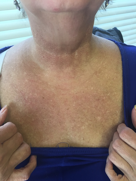
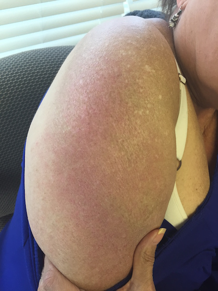
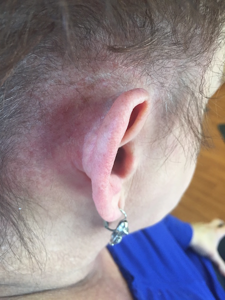
CLINICAL HISTORY: The patient is a 64-year-old female with hypertension, hyperlipidemia, hypercholesterolemia, hypothyroid, and depression who presented to the clinic with a pruritic rash that started on her left wrist and then spread to her right arm, chest, scalp, and posterior neck. She denied any recent sun exposure. The patient admits to some difficulty arising from a seated position as well as fatigue while combing her hair. The patient had been given multiple topical steroids with only minimal relief of the rash and the associated pruritus. Lab work, muscle, and skin biopsy were ordered, as well as follow up with rheumatology in regards to a muscle biopsy. The patient had been to multiple physicians prior to coming to our clinic including an internal medicine physician, dermatologist, allergist, rheumatologist, as well as her primary care physician. The patient admits having all normal screening exams such as a mammogram/colonoscopy/pelvic examination as well as a recent CT of her chest, abdomen, and pelvis which didn’t reveal any abnormalities.
Medications: amlodipine, aspirin, clopidogrel, enalapril, hydralazine, hydrochlorothiazide, metoprolol, nitroglycerin prn, duloxetine, atorvastatin
PHYSICAL EXAM:
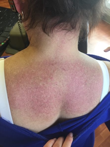
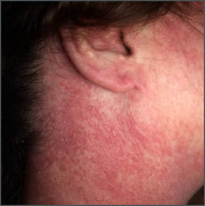
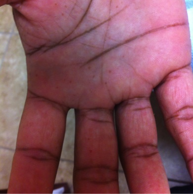
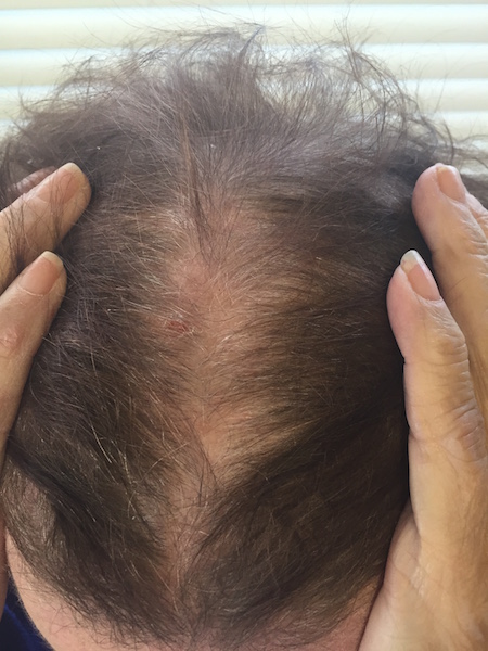
Well-nourished, healthy appearing female with scaly pink erythematous patches on bilateral upper extremities, post-auricular bilaterally, central chest, and anterior/posterior neck. The patient also had scaly erythema along frontal forehead near her hairline, and violaceous poikiloderma especially of the lateral neck, superior back/neck, and chest. She stated previously on her hands she had erythematous papules overlying the metacarpophalangeal joints but this has since resolved.
LABORATORY TESTS:
ALT-53, AST- 46, Aldolase-10.4, CPK-297.1, WBC-2.8, Platelets-122
DERMATOHISTOPATHOLOGY:
Skin biopsy demonstrated epidermal atrophy, vacuolar interface change, mucin deposition, and a heavy lymphocytic infiltrate. Muscle biopsy showed perifascicular atrophy of myofibers as well as capillary depletion. Also, the biopsy demonstrated a CD4+ lymphocytic infiltrate.
DIFFERENTIAL DIAGNOSIS:
1. Mixed Connective Tissue Disease
2. Dermatomyositis
3. Systemic Lupus Erythematosus
4. Photoallergic Drug
5. Polymorphus Light Eruption

