CORRECT DIAGNOSIS:
Dermatomyositis
DISCUSSION:
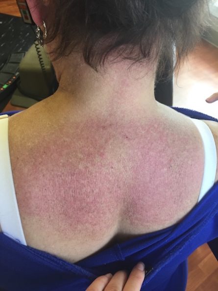
Dermatomyositis (DM) is a chronic multisystem autoimmune connective tissue disease. Dermatomyositis has a bimodal distribution (10-15 years old) and (45-60 years old). DM can be skin only (amyopathic) or only muscle (polymyositis) or involve both entities. The adult form is generally 2:1 female to male ratio. The skin findings usually present 2-3 months prior to muscle involvement. It presents clinically with skin findings of facial erythema, violaceous papules overlying the metacarpophalangeal joints (Gottron papules), erythematous scaly plaques over extensor surfaces such as elbows, knees, and knuckles, ragged cuticles (samitz sign) with increased nail fold telangiectasis and drop out of capillaries, and photodistributed poikiloderma including of the eyelids with edema (heliotrope rash). Other features include an erythematous patch over posterior neck and back (shawl sign) as well as of the anterior chest. Patients may also have poikiloderma over lateral hip (holster sign). Patients can also have calcinosis cutis which is seen more commonly in juvenile patients. Pruritus is commonly associated with the rash. Muscle involvement is seen clinically as proximal muscle weakness such as the inability to comb one’s hair or arise from the seated position. Up to 40% of adult patients may have an associated occult malignancy which includes in men most commonly (lung and GI carcinoma) and women (ovarian and breast). The diagnosis is made on a combination of clinical history/presentation, histologic findings (which look similar to lupus erythematosus), and/or a muscle biopsy or MRI. Laboratory findings may include a positive ANA (60%), increased creatinine kinase (90%), aldolase, ESR, LDH, and transaminases. The treatment can include oral corticosteroids followed by a steroid-sparing agent, antimalarial, and sun protection. A recent study by Kurtzman et al. found tofacitinib citrate to be successful in treating recalcitrant dermatomyositis, which could be of interest to our patient.
TREATMENT:
Our patient was started on Imuran 50 mg BID which has been well-tolerated by her. She is very good about sun protection and has been receiving intralesional injections of Kenalog 10mg/cc into her scalp every month to help with the pruritus and scale. She has also been started on marinol 2.5 mg BID to help with the pruritic rash. The patient was also switched to a different lipid-lowering agent as she was initially placed on atorvastatin, which has been reported in the literature to worsen DM. She has also received all recommended imaging/screening studies in regard to the increased cancer propensity with DM including a pelvic examination, colonoscopy, mammogram, and a CT of her chest/abdomen/pelvis all within the last year. No abnormalities were found.
REFERENCES:
Bolognia, J. L., Jorizzo, J. L., & Schaffer, J. V. (2012). Dermatology (3rd ed., pp. 159-161). Spain: Elsevier.
Elston, D. M., & Ferringer, T. (2014). Dermatopathology (2nd ed., p. 141). N.p.: Saunders Elsevier.
Kurtzman, D. B., Wright, N. A., Lin, J., Femia, A. N., Merola, J. F., Patel, M., & Vleugels, R. A. (2016). Tofacitinib citrate for refractory cutaneous dermatomyositis. JAMA Dermatology. https://doi.org/10.1001/jamadermatol.2016.0661
Thual, N., Penven, K., Chevallier, J. M., Dompmartin, A., & Leroy, D. (2005). Fluvastatin-induced dermatomyositis. Annales de Dermatologie et de Vénéréologie, 132(12 Pt 1), 996-999.


