Presenter: Leslie Mills, DO
Dermatology Program: West Palm Hospital/PBCGME
Program Director: Robin Shecter, DO
Submitted on: May 2, 2016
CHIEF COMPLAINT: Painful rash involving her face, neck, and ears
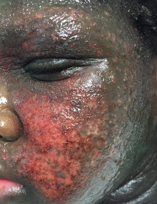
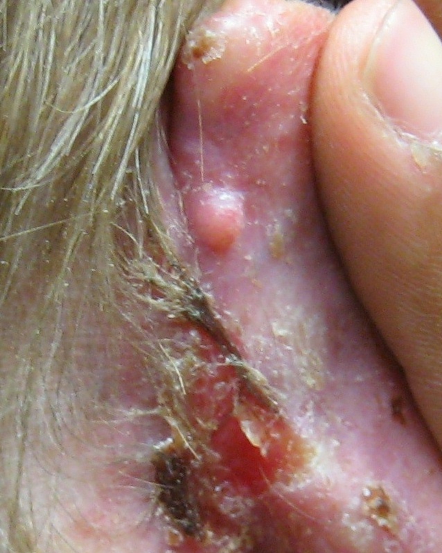
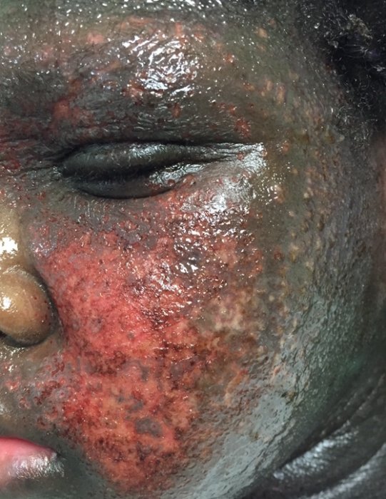
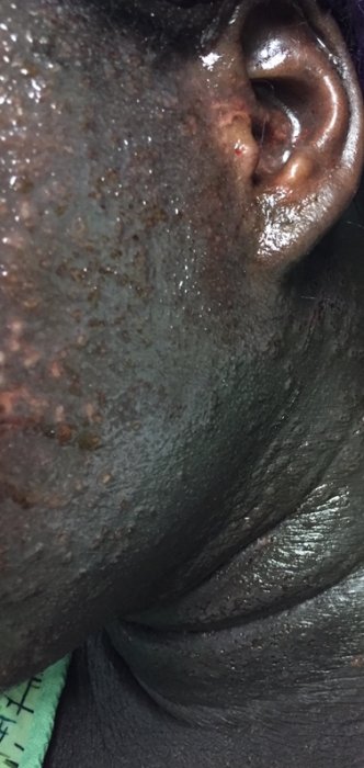
CLINICAL HISTORY: A 15-year-old female presented to the Emergency Department with a painful rash affecting her face, neck, and ears. Four days prior to admission, after returning from Georgia, she experienced tenderness and pressure in her facial area, which progressed to significant edema, particularly in the periorbital region. The rash initially appeared as intensely pruritic, erythematous papules and vesicles that rapidly ulcerated, producing clear-yellow drainage. The patient reported associated ocular pain but denied experiencing oral lesions, fever, or chills. She also had no recent trauma, sick contacts, or exposure to animals, nor had she engaged in outdoor activities or used new products.
The patient had no previous treatment for this current episode, having recently discontinued prednisone and triamcinolone ointment for a similar rash that previously affected her lower extremities. Her past medical history is notable for eczema and food and environmental allergies. Family history includes a brother with eczema and asthma. Socially, she lives with her parents, attends high school, and denies any use of tobacco, alcohol, or illicit drugs. Her surgical history is unremarkable, and her current medication regimen includes Zyrtec, with no known drug allergies.
PHYSICAL EXAM:
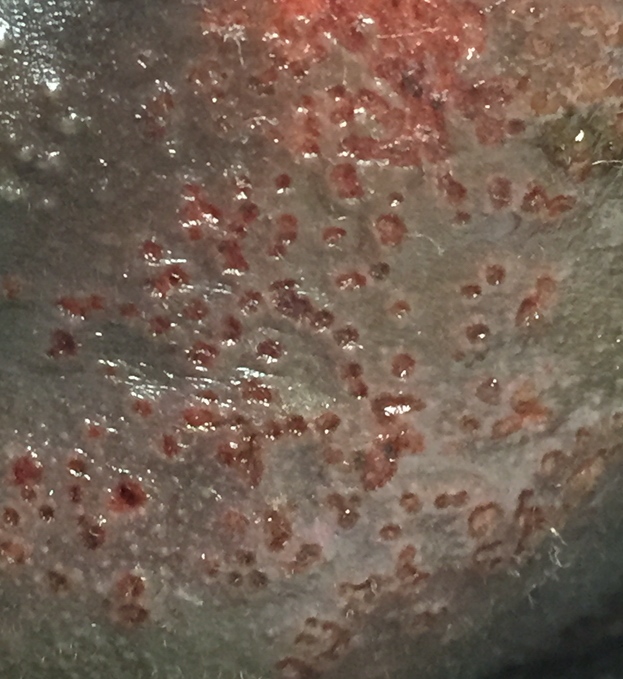
Multiple, tender, discrete, 2-3mm punched-out erosions with hemorrhagic crusts primarily involving the face, neck, and ears with clear-yellow drainage, erythema, and significant periorbital edema.
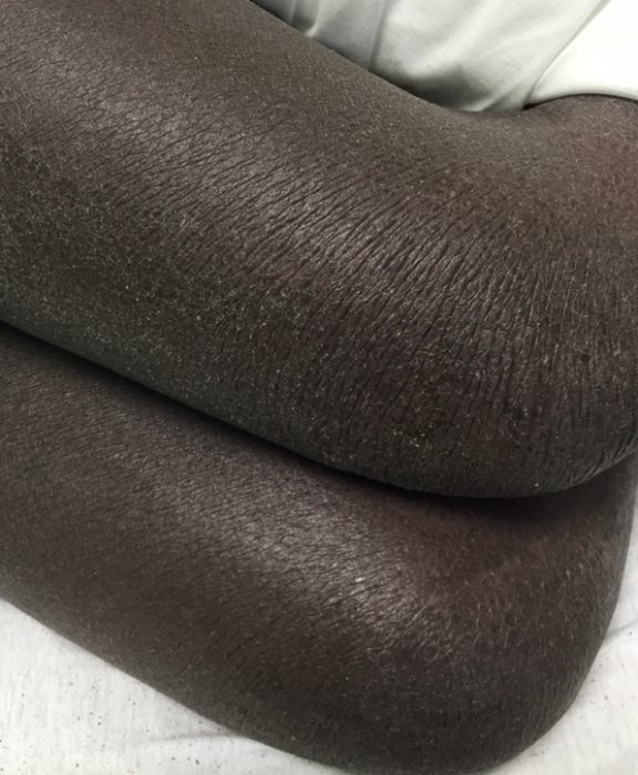
Associated physical findings included hyperlinear palms and thickened, dry, scaly plaques involving the face, neck, flexural surfaces, and extremities.
LABORATORY TESTS:
HSV-1 DNA PCR – positive
HSV-2 DNA PCR – negative
Viral culture – herpes simplex virus
Bacterial culture – Staphylococcus aureus
DERMATOHISTOPATHOLOGY:
Epidermal necrosis, enlarged epithelial cells with intranuclear inclusions, and multinucleated giant cells with polymorphonuclear leukocyte invasion of the epidermis and dermis.
DIFFERENTIAL DIAGNOSIS:
1. Varicella zoster virus infection
2. Bullous impetigo
3. Erysipelas
4. Contact dermatitis
5. Bullous drug eruption

