CORRECT DIAGNOSIS:
Eczema Herpeticum
DISCUSSION:
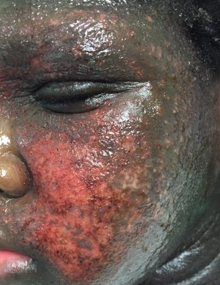
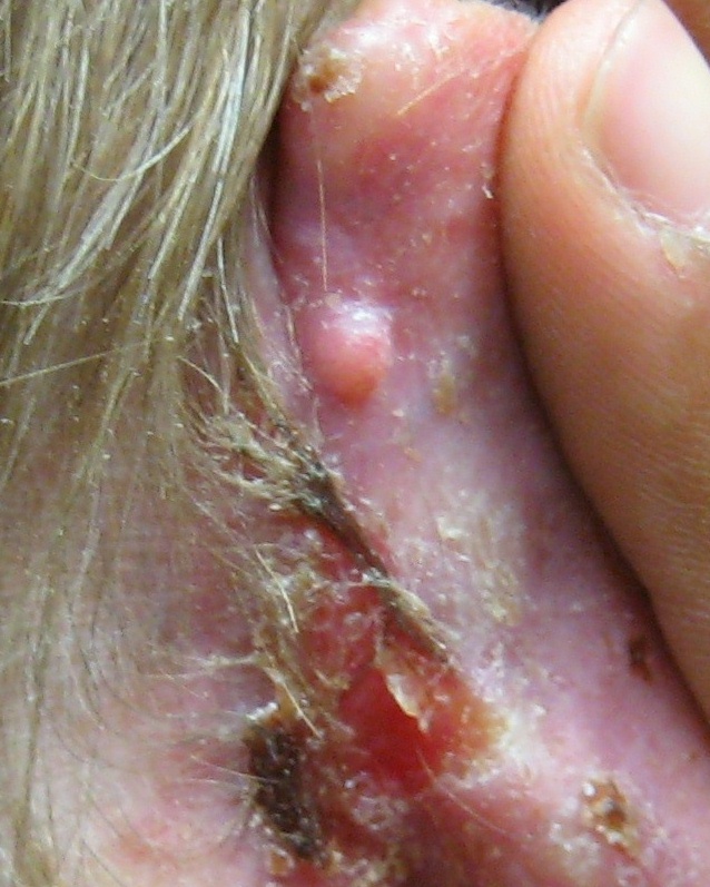
Clinically, our patient presented with features highly suspicious for eczema herpeticum (EH) or Kaposi’s varicelliform eruption (KVE), a diagnosis later confirmed by laboratory studies.
Eczema herpeticum represents rapid dissemination of herpes simplex viral (HSV) infection occurring on eczematous dermatitis, most often due to HSV-1 infection in infants and children with atopic dermatitis. Patients with mutations in the filaggrin gene and those who have severe atopic dermatitis and asthma are at an increased risk for EH. Higher IgE levels, food and environmental allergies, and peripheral eosinophilia may also be associated findings. Bath or hot tub exposure has been reported as a risk factor as well.
A decreased production of antimicrobial peptides in the epidermis represents an important defense against cutaneous HSV infection. Increased IL-10-producing monocytes lead to local expansion of regulatory T-cells and may contribute to the development of EH. HLA-B7 and IL-25 expression may also be noted.
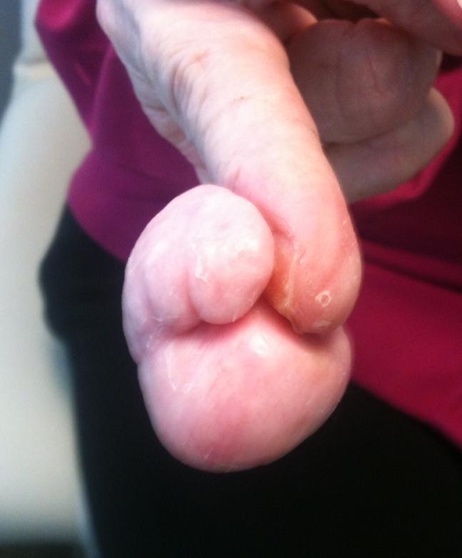
Cutaneous dissemination of HSV may also occur in the setting of ichthyoses (highly suspected in our case), severe seborrheic dermatitis, scabies, Darier’s disease, benign familial pemphigus, pemphigus (foliaceus or vulgaris), pemphigoid, cutaneous T-cell lymphoma, Wiskott-Aldrich syndrome, allergic or photoallergic contact dermatitis, and burns.
Eczema herpeticum initially develops as an eruption of vesicles but affected individuals more often present with numerous monomorphic, punched-out erosions with hemorrhagic crusting as demonstrated by our patient. Eczema herpeticum is frequently widespread and may occur at any site, a feature also reported by our patient who described similar lesions involving her lower extremities just weeks prior to admission. Patients may have fevers, malaise, and lymphadenopathy. Lesions may be complicated by bacterial superinfections, most commonly Staphylococcus aureus and/or Streptococcus pyogenes. Additional complications may include herpetic keratoconjunctivitis and meningoencephalitis.
Multiple laboratory tests are available to diagnose HSV infection. In addition to a Tzanck smear, DFA is frequently performed as an initial diagnostic test due to its greater sensitivity, ability to distinguish between HSV and VZV, and rapid turnaround time. The identification of HSV by viral culture usually requires 2-5 days. PCR is the preferred method for identifying HSV in the cerebrospinal fluid and is increasingly being used as a rapid, sensitive, and specific method to detect HSV DNA in tissue.
Histologic examination reveals enlarged, slate-gray keratinocyte nuclei with margination of chromatin, followed by intraepidermal vesiculation associated with ballooning degeneration of keratinocytes. These swollen, pale keratinocytes often fuse to form multinucleated giant cells and may contain eosinophilic intranuclear inclusion bodies surrounded by an artifactual cleft (Cowdry type A inclusions) affecting adnexal structures and interfollicular epidermis. A variable dense dermal infiltrate of lymphocytes, neutrophils, and eosinophils are observed. Vascular changes may include areas of hemorrhagic necrosis of the epidermis.
TREATMENT:
Due to the potential seriousness of this disorder, treatment with systemic antiviral agents is indicated. Hospitalization with the administration of intravenous acyclovir or foscarnet (in acyclovir-resistant cases) may be required. In stable patients with less severe disease, oral valacyclovir or famciclovir (both prodrugs of acyclovir) may be considered. If hospitalized, patients should be isolated from other patients with universal protective precautions. Maximize supportive measures in systemically ill patients, including fluid and electrolyte imbalances, wound care, pain control, and nutrition. Monitor and promptly treat any complicating infection with the appropriate antibiotic therapy based on culture sensitivities. Symptomatic relief may be accomplished with cool compresses and emollients.
REFERENCES:
Bolognia, J., Jorizzo, J., Schaffer, J., et al. (2012). Dermatology (3rd ed.). Amsterdam, Netherlands: Elsevier. pp. 211, 1321-1327.
Bussmann, C., Peng, W. M., Bieber, T., & Novak, N. (2008). Molecular pathogenesis and clinical implications of eczema herpeticum. Expert Review of Molecular Medicine, 10, e21. https://doi.org/10.1017/S1462399408000418. PubMed ID: 18620613.
Du Vivier, A. (2013). Recurrent herpes simplex. In Atlas of Clinical Dermatology (4th ed., pp. 307-311). Philadelphia, PA: Elsevier.
James, W. D., Berger, T. G., Elston, D. M., et al. (2016). Viral diseases. In Andrews’ Diseases of the Skin (12th ed., pp. 365-367). Philadelphia, PA: Elsevier.
Kramer, S. C., Thomas, C. J., Tyler, W. B., & Elston, D. M. (2004). Kaposi’s varicelliform eruption: A case report and review of the literature. Cutis, 73(2), 115-122. PubMed ID: 15027517.
Marques, A. R., & Cohen, J. I. (2012). Herpes simplex. In Fitzpatrick’s Dermatology in General Medicine (8th ed., pp. 2367-2382). New York, NY: McGraw-Hill.


