CORRECT DIAGNOSIS:
Neurofibromatosis type 1
DISCUSSION:
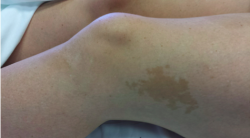
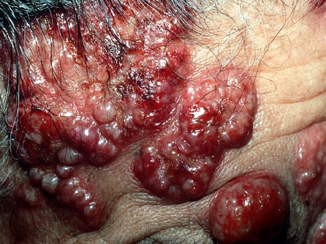
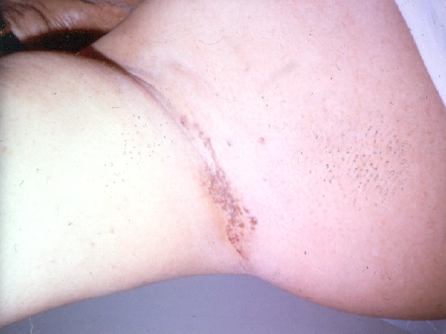
Neurofibromatosis type 1 also known as Von Recklinghausen Disease is an autosomal dominant condition occurring in 1 of 3000 patients. The pathophysiology includes loss of function of the tumor suppressor NF1 on chromosome 17q11. Clinical findings include a spectrum of variation with cutaneous, ocular, skeletal, neurological, and cardiac manifestations. Diagnostic criteria include two or more of the following: six or more cafe au lait macules greater than 5mm in prepubertal individuals and greater than 15 mm in adults, two or more neurofibromas (NF) or one plexiform, axillary or inguinal freckling, optic gliomas, two or more lisch nodules, osseous lesions such as sphenoid wing dysplasia or thinning of long bones, and a first degree relative with NF1. Axillary freckling also known as Crowe’s sign is the most specific finding and a plexiform NF is pathognomonic for Neurofibromatosis type 1.
TREATMENT:
Genetic counseling and monitoring are recommended. Ophthalmology consultation was performed as well.
Malignant transformation can occur and observation must be made for the following tumors: optic nerve glioma, malignant peripheral nerve sheath tumor, GI stromal tumors, and pheochromocytoma.
In conclusion, we present a new diagnosis of Neurofibromatosis type 1 in a 17-year-old girl. Minimum annual evaluation for adults with the uncomplicated disease will include a dermatologic exam, MRI and neurological evaluation, blood pressure measurement, and age-appropriate cancer screenings. Potentially affected family members will require a cutaneous and ophthalmological exam with genetic testing as a consideration.
REFERENCES:
Blakeley, J. O., & Plotkin, S. R. (2016). Therapeutic advances for the tumors associated with neurofibromatosis type 1, type 2, and schwannomatosis. Neuro-Oncology, 18(2), 124-136. https://doi.org/10.1093/neuonc/nov200
Bolognia, J. (2012). Neurofibromatosis and Tuberous Sclerosis. In Dermatology (3rd ed., Chapter 61). Elsevier Saunders.
Tekin, M., Bodurtha, J. N., & Riccardi, V. M. (2001). Café au lait spots: The pediatrician’s perspective. Pediatrics in Review, 22(3), 82-90. https://doi.org/10.1542/pir.22-3-82
Louprasong, A., & Mercado, K. J. (2016). Ocular signs of neurofibromatosis. Retrieved May 31, 2016, from https://www.reviewofoptometry.com/article/ocular-signs-of-neurofibromatosis
Shah, K. N. (2010). The diagnostic and clinical significance of café-au-lait macules. Pediatric Clinics of North America, 57(5), 1131-1153. https://doi.org/10.1016/j.pcl.2010.07.002
Takahasi, M. (1976). Studies on café au lait spots in neurofibromatosis and pigmented macules of nevus spilus. Tohoku Journal of Experimental Medicine, 118(3), 255-273. https://doi.org/10.1620/tjem.118.255
Urganci, N., Genc, D. B., Kose, G., Onal, Z., & Vidin, O. O. (2015). Colorectal cancer due to constitutional mismatch repair deficiency mimicking neurofibromatosis I. Pediatrics, 136(4), e1047-1050. https://doi.org/10.1542/peds.2015-1426
Ma, D. L., & Hu, J. (2015). Images in clinical medicine: Segmental neurofibromatosis. New England Journal of Medicine, 372(10), 963. https://doi.org/10.1056/NEJMicm1403193
Hernández-Martín, A., & Duat-Rodríguez, A. (2016). An update on neurofibromatosis type 1: Not just café-au-lait spots and freckling. Part II. Other skin manifestations characteristic of NF1. NF1 and cancer. Actas Dermosifiliogr, 107(3), 234-240. https://doi.org/10.1016/j.ad.2016.01.009


