CORRECT DIAGNOSIS:
Radiation-Induced morphea
DISCUSSION:
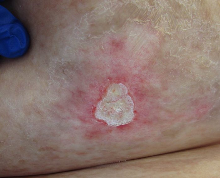
Radiation-induced morphea (RIM) is a rare complication of radiotherapy. Less than 100 cases have been reported in the literature, with the majority occurring in women treated for breast carcinoma. The frequency of RIM of the breast is 0.2%. No correlation has been found between the incidence of RIM and age, total dose of radiation, dose per fraction, or acute reaction severity. RIM has a non-characteristic appearance and often goes unrecognized or misdiagnosed.
Radiation therapy causes cutaneous reactions in more than 90% of patients. These reactions can be divided into early and late side effects. Early side effects occur three to six weeks post-radiation and include epilation, hyperpigmentation, desquamation, and erythema. Late-stage changes include hyperpigmentation, fibrosis, and telangiectasia and can occur anywhere from six weeks to three years post-radiation. RIM is a late side effect that commonly presents one to three years after therapy. It is often painful and has an initial inflammatory phase followed by a sclerotic phase characterized by induration, fibrotic retraction, and pigmentation of the breast.
The pathogenesis of RIM is theorized to be a T-cell reaction to radiation-induced neoantigens. The newly formed antigens stimulate the secretion of cytokines including transforming growth factor-β (TGF-β), a potent inducer of collagen synthesis. The pathologic secretion of TGF-β causes extensive fibrosis, ultimately leading to localized morphea.
Histopathologically, the inflammatory phase is characterized by an interstitial lymphoplasmacytic infiltrate that can include eosinophils among slightly thickened collagen bundles. Areas of subcutaneous fat are replaced by newly formed collagen, and vascular changes consist of endothelial swelling. In the sclerotic phase, collagen bundles in the reticular dermis are thickened and closely packed together. An inflammatory infiltrate is often absent, except in some areas of the subcutis.
The differential diagnosis of RIM includes tumor recurrence, acute and chronic radiation dermatitis, post-irradiation fibrosis (PIF), and cellulitis. Acute radiation dermatitis is characterized histologically by edema, vasodilation, and erythrocyte extravasation. Chronic radiation dermatitis can be differentiated by the presence of atypical fibroblasts, which are not seen in RIM. PIF is a more common entity characterized by deep subcutaneous and fascial fibrosis with little to no inflammatory infiltrate compared to the primarily dermal fibrosis seen with RIM.
RIM is a difficult condition to treat. Some cases spontaneously improve, but the majority are unresponsive to therapy. Topical and systemic steroids, oral antibiotics, systemic immunosuppressants, topical imiquimod, and topical vitamin D derivatives have all been used with varying degrees of success. PUVA therapy has been reported to cause skin softening but does not reverse fibrosis and atrophy, therefore prompt initiation of treatment is important to achieve the best possible outcome.
TREATMENT:
The patient’s therapeutic regimen includes intralesional corticosteroid injections and topical clobetasol cream. She is also using an aromatase inhibitor as adjuvant therapy for breast carcinoma.
REFERENCES:
Cheah, N., Wong, D., & Chetiyawardana, A. (2008). Radiation-induced morphea of the breast: A case report. Journal of Medical Case Reports, 2(1), 136. https://doi.org/10.1186/1752-1947-2-136
Davis, D., Cohen, P., McNeese, M., et al. (1996). Localized scleroderma in breast cancer patients treated with supervoltage external beam radiation: Radiation port scleroderma. Journal of the American Academy of Dermatology, 35(6), 923-927. https://doi.org/10.1016/S0190-9622(96)90381-7
Dubner, S., Bovi, J., White, J., et al. (2006). Postirradiation morphea in a breast cancer patient. Breast Journal, 12(2), 173-176. https://doi.org/10.1111/j.1075-122X.2006.00238.x
Nakagawa, H. (2015). A case of radiation-induced generalized morphea with prominent mucin deposition and tenderness. American Journal of Case Reports, 16, 279-282. https://doi.org/10.12659/AJCR.894445
Spalek, M., Jonska-Gmyrek, J., & Gałecki, J. (2014). Radiation-induced morphea: A literature review. Journal of the European Academy of Dermatology and Venereology, 29(2), 197-202. https://doi.org/10.1111/j.1468-3083.2013.04514.x


