CORRECT DIAGNOSIS:
Metastatic Breast Carcinoma
DISCUSSION:
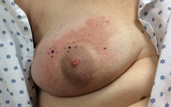
The most frequently diagnosed cancer in females is breast carcinoma.1,2 Up to 10% of breast cancer cases present with metastasis, which is typically a recurrence of early-stage breast carcinoma. Breast cancer is the most common cancer to involve the skin with cutaneous involvement being the presenting sign in 3.5% of cases.1 The most common site of cutaneous metastases of breast cancer in the chest wall, as well as the abdomen and scalp. Up to eighty percent of cases present with non-tender, firm to rubbery nodules.1,3 There are several other specific cutaneous morphologic patterns of metastatic breast carcinoma including carcinoma en cuirasse, carcinoma telangiectaticum, carcinoma erysipeloid and alopecia neoplastica. Carcinoma en cuirasse, also referred to as scirrhous carcinoma, presents as a diffuse, indurated erythematous plaque with a leathery or fibrotic texture on the chest wall. Carcinoma telangiectaticum presents with pruritic pink to erythematous to violaceous papules, plaques, or nodules with telangiectasia.1,3 Carcinoma Erysipeloid, also known as inflammatory metastatic carcinoma, presents as tender, well-defined erythematous edematous warm plaques. Alopecia neoplastica presents as well-demarcated annular plaques of cicatricial alopecia on the scalp.1,3 The above morphologic patterns are not specific for, but are highly suggestive of, metastatic breast carcinoma as they can also be seen in metastatic tumors of varying origins. Breast carcinoma can also present as a paraneoplastic syndrome including erythema gyratum repens, acquired ichthyosis, dermatomyositis, multicentric reticulohistiocytosis, and hypertrichosis lanuginosa acquisita.1 Identifying these various cutaneous presentations is of the utmost importance as Dermatologists, as we can contribute to the diagnosis of unrecognized cancers and assist in prompt referral to the appropriate specialist for further care.
In our patient’s case, she had a past medical and surgical history consistent with breast carcinoma in the same breast prior to the onset of her rash which helped to immediately raise our index of suspicion for metastatic breast carcinoma. Not all cases have that prior history and it can sometimes be challenging to distinguish between some of the benign and malignant causes of nipple dermatitis. Malignant causes are typically unilateral and present in some of the ways discussed above. Mammary Paget’s disease (MPD) should always be in the differential diagnosis of unilateral nipple dermatitis. Once MPD is diagnosed, it is associated with underlying breast cancer in 93-100% of cases.4 It presents as a unilateral erythematous, eczematous patch to plaque on the breast with an overlying scale which can be pruritic and crusted or lead to ulceration. In comparison to the histopathology of metastatic breast carcinoma, Paget’s disease will affect the epidermis showing enlarged cells with atypical, pleomorphic nuclei surrounded by increased cytoplasm. These cells show upward migration in the epidermis and stain positively for cytokeratin-7 and carcinoembryonic antigen.4 Other causes of nipple dermatitis can involve benign etiologies including atopic dermatitis, allergic or irritant contact dermatitis and infectious causes. Nipple dermatitis can be seen in 12-23% of patients with atopic dermatitis. It commonly presents as bilateral erythematous eczematous patches with serum crust with or without vesicles on the areola that extend onto the periareolar skin and will be responsive to topical corticosteroids.3,5 Irritant contact dermatitis is classically seen on the bilateral nipples, as well as the areola, due to frictional causes including ill-fitting brassieres in women or irritation from shirts in joggers. Allergic contact dermatitis can be due to allergens applied to the nipple or breast directly, a common finding in mother’s trying to relieve the pain or soreness from breastfeeding.3 Secondary infectious causes are also commonly present in breastfeeding mothers. Candidiasis of the nipple is characterized by a red to violaceous discoloration of the nipple and proximal areola as well as severe burning pain which radiates throughout the breast which is worse after feedings.6 Factors that predispose breastfeeding mothers to nipple candidiasis include recent vaginal candidiasis, cracked or traumatized nipples, and recent antibiotic use. If nipple candidiasis is suspected, the infant should be evaluated for both oral thrush and diaper candidiasis.6
Although nipple dermatitis can be a challenging problem as a practitioner, a thorough history and physical examination should help to narrow the differential diagnoses. Any unilateral eczematous rash on the breast which has been unresponsive to appropriate treatment measures and persisted for longer than three months requires a biopsy to rule out malignant etiologies.3
TREATMENT:
The patient was subsequently referred to the Oncology Department of a tertiary medical center for complete workup and staging.
REFERENCES:
1. Tan AR. Cutaneous manifestations of breast cancer. Seminars in Oncology. 43 (2016) 331-334
2. Marcu A, Lyratzopoulos G, Black G, et al. Educational differenes in likelihood of attributing breast symptoms to cancer: a vignette-based study. Psycho-Oncology. 17 May 2016.
3. James WD, Berger TG, Elston EM. Andrews’ Diseases of the Skin Clinical Dermatology. Eleventh Edition. Elsevier Publishing 2011. Pgs 72, 616
4. Filho LLL, Lopes LRS, Michalany AO, et al. Mammary and Extramammary Paget’s disease. An Brs Dermatol. 2015; 90(2):225-31.
5. Pugliarello S, Cozzi A, Gisondi P, et al. Phenotypes of atopic dermatitis. JDDG; 2011. 9:12-20.
6. Tanguay KE, McBean MR, Jain E. Nipple candidiasis among breast feeding mothers. Can Fam Physician. 1994; 40:1407-1413.


