CORRECT DIAGNOSIS:
Leprosy
DISCUSSION:
Leprosy, or Hansen’s disease, is a chronic infectious disease with prominent involvement of the skin and nerves. It is caused by Mycobacterium leprae with descriptions of infection dating back as early as 600B.C. Leprosy is rarely fatal, but it does represent a disabling, deforming, stigmatizing disease. It was once a worldwide health issue. Currently, it has been substantially reduced to a prevalence rate of less than 1 case per 10,000 people in the majority of the world. 95% of cases now occur in 16 countries in regions of Asia, Africa, Central and South America where there seems to be a lower standard of living and poorer hygiene. There were, however, 168 cases diagnosed in the United States in 2012. They were endemic in the southeast coastal regions and Hawaii. Transmission typically occurs after contact with an infected individual, or in the case of the U.S., contact with the 9-banded armadillo. It is estimated that > 90% of exposed persons are resistant to M. leprae, and 1.7-31% of the population in endemic areas are seropositive for specific antigens. Early diagnosis and prompt treatment are vital to control leprosy, but are often delayed. Leprosy is not typically suspected in developed countries. In fact, in the U.S. diagnosis is delayed on average by 1 ½ -2 years. In addition, identification of the organism in the affected tissue is difficult as M. leprae cannot be cultured. To aid in diagnosing leprosy, biopsies with fite acid-fast stain (developed world) as well as identifying the organisms on slit smear of skin (developing world) can be performed. Unfortunately, serologic tests and PCR to detect small numbers of organisms have not improved diagnosis thus far.
Leprosy can be divided into two major forms on the extremes of the spectrum, tuberculoid or lepromatous, which depends on the degree and type of immunity. Several intermediate forms are located along the spectrum. In indeterminate leprosy, the earliest lesion is typically a solitary, hypopigmented, anesthetic macule most commonly located on the cheeks, upper arms, thighs, or buttocks. Although the classification is indeterminate, the diagnosis is not, as it may “progress” to anywhere on the spectrum. Tuberculoid leprosy is relatively stable and typically evolves slowly and often spontaneously remits in about 3 years. 1-5 asymmetric lesions that are hypopigmented or erythematous dry, scaly, anesthetic plaques are present. Additionally, enlarged nerves (greater auricular and superficial peroneal may be visible). Borderline tuberculoid leprosy resembles tuberculoid lesions, but are smaller and more numerous, sometimes with characteristic satellite lesions around larger lesions. Numerous (but countable) red, irregularly shaped plaques, with enlarged, tender nerves, and only moderate anesthesia of plaques are seen in borderline leprosy.
Borderline lepromatous leprosy lesions are symmetric and too numerous to count. They consist of macules, papules, plaques, and nodules. Sensation and sweating are normal with nerve involvement occurring later.
In lepromatous leprosy, symmetric, hypopigmented macules are evident with minimal or no loss of sensation, no nerve thickening, or change in sweating. However, nerve involvement develops very slowly and is symmetric, typically in a stocking-glove distribution. This often leads to a misdiagnosis of diabetic neuropathy.
TREATMENT:
The management of leprosy is based on bacterial load (paucibacillary vs multibacillary) with more than 5 lesions or bacilli visible on smear or biopsy considered multibacillary. According to the US Public Health Service recommendations, paucibacillary infections should be treated with 600mg rifampin and 100mg dapsone daily for 1 year and multibacillary infections require 600mg rifampin, 100mg dapsone, and 50mg clofazimine daily for 2 years. It is noted that 100mg minocycline or 400mg ofloxacin may be substituted for clofazimine if warranted.
Additional Comment:
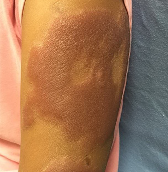
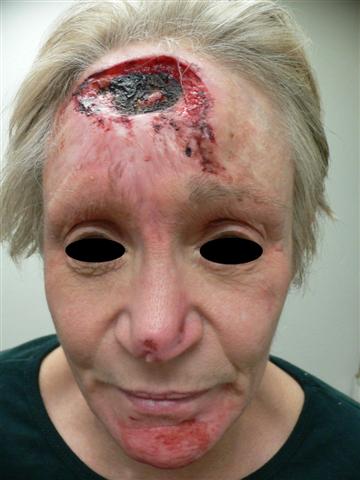
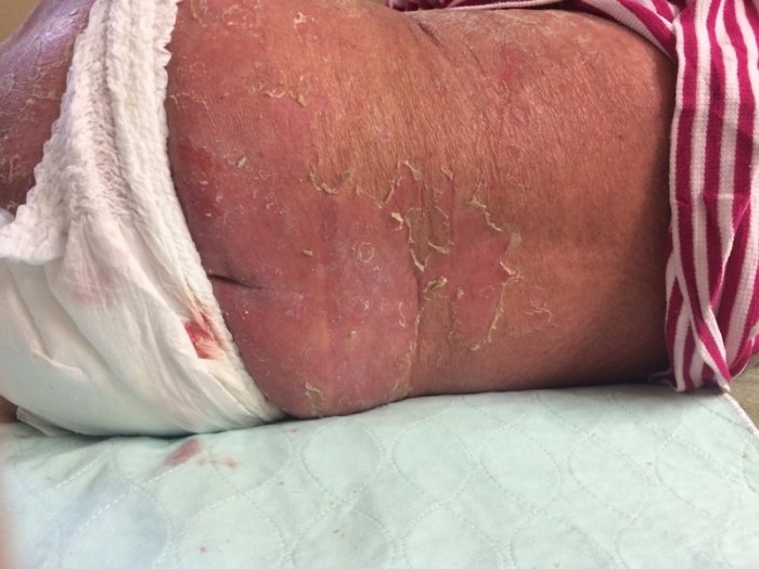
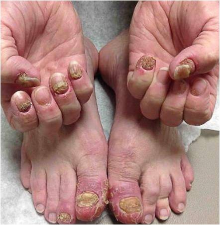
Our patient had an examination most concerning for tuberculoid leprosy in the setting of erythematous to brown indurated plaques of the face, extremities, and less involvement of the trunk. Tuberculoid leprosy does lead to anesthetic skin lesions due to damage to superficial peripheral nerves. However, she had features of both tuberculoid and lepromatous leprosy, which is likely consistent with a borderline presentation (possibly from a previous treatment). She also had neuropathic ulceration of the left first metatarsal bone as well as anesthesia to indurated plaques of the lower extremities. Her upper extremity plaques are newer with no decreased sensation on examination but more hypoesthetic than what would be expected (also can be seen as well). Lastly, the patient also moved from an endemic area known for tuberculoid leprosy. The physical findings in conjunction with all positive results led us to a diagnosis of leprosy. The patient was started on a regimen of minocycline 100mg bid, dapsone 100mg qd, rifampin 600mg qd, and prednisone 60mg per infectious disease. She was referred to the official Hansen treatment center for definitive care.
Newer drug regimens suggested for leprosy in 2009 by the “WHO Report of the Global Programm Managers’ Meeting on Leprosy Control Strategy” is as follows: for rifampicin susceptible MB patients a fully supervised monthly regimen could include: Rifapentin 900 mg (or rifampicin 600 mg), moxifloxacin 400 mg, and clarithromycin 1000 mg (or minocycline 200 mg) for 12 months. For rifampicin-resistant patients, the intensive phase could include moxifloxacin 400 mg, clofazimine 50 mg, clarithromycin 500 mg, and minocycline 100 mg daily supervised for six months. The continuation phase could comprise moxifloxacin 400 mg, clarithromycin 1000 mg, and minocycline 200 mg once monthly, supervised for an additional 18 months
REFERENCES:
Andrews’ Diseases of the Skin.
Firooz, A. (2006). Multidrug therapy regimen for leprosy. Journal of the American Academy of Dermatology, 55(6), 1115.
Health Resources & Services Administration. U.S. Department of Health and Human Services.
Moschella, S. L. (2004). An update on the diagnosis and treatment of leprosy. Journal of the American Academy of Dermatology, 51(3), 417–426.
Rao, P. N., & Jain, S. (2013). Newer management options in leprosy. Indian Journal of Dermatology, 58(1), 6-11.
Silva, M. R., & Castro, M. C. R. (2012). Mycobacterial infections. In J. L. Bolognia, J. L. Jorizzo, & J. V. Schaffer (Eds.), Dermatology (Vol. 2, pp. 1221-1228). Amsterdam: Elsevier Limited.


