Presenter: Richard Winkelmann, DO; Jessica Hoy, DO; Kylee Sacksteder, DO; Gabriella Maloney, DO; Alyson Ridpath, DO
Dermatology Program: OhioHealth Dermatology Columbus, OH
Program Director: Dawn Sammons, DO
Submitted on: May 5, 2017
CHIEF COMPLAINT: Recurrent bumps on back of right lower leg
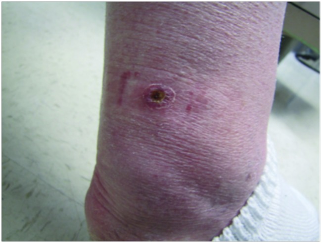
CLINICAL HISTORY: A 64-year-old immunocompetent female presented with a seven-month history of recurrent erythematous papules and nodules on the back of her right lower leg. She reported that the nodules were tender, nonpruritic, and, at times, had a clear exudate. The patient denied any trauma to the area and initially attributed the eruption to mosquito bites. No previous treatments. Patient denied any personal or family history of skin cancers, and her medical history was unremarkable without prior exposure to tuberculosis or recent travel out of the country.
PHYSICAL EXAM:
Clinical examination revealed three 5 to 6mm inflamed, subcutaneous, violaceous nodules with a fine overlying scale on the posterior aspect of the right lower leg. Lesions were surrounded by several hyperpigmented macules at sites of previous lesions.
LABORATORY TESTS:
CBC and CMP evaluations were within normal limits. Chest x-ray showed no evidence of active or latent pulmonary tuberculosis and PPD testing was negative. The patient’s hepatitis panel was normal with no evidence of prior infection.
DERMATOHISTOPATHOLOGY:
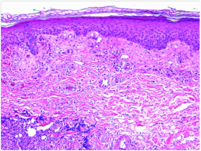
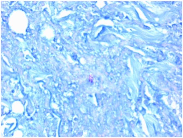
A 4mm punch biopsy of the lesion to the level of the dermis taken from the right posterior lower leg revealed suppurative inflammation and foci of necrosis. Acid-fast staining demonstrated acid fast, beaded bacilli consistent with mycobacterial infection. Another 3mm punch biopsy of the lesion from the right posterior lower leg was performed for tissue culture. The culture identified Mycobacterium chelonei as the pathogenic organism.
DIFFERENTIAL DIAGNOSIS:
1. Erythema induratum
2. Erythema nodosum
3. Panniculitis
4. Polyarteritis nodosa
5. Atypical Mycobacterium


