Presenter: Victoria Comeau, D.O., Alexandria Glass, D.O., Caitlin Porubsky, D.O., Carmen Julian, D.O., Marcus Goodman, D.O.
Dermatology Program: PCOM Dermatology Residency
Program Director: Marcus Goodman, D.O.
Submitted on: September 7, 2017
CHIEF COMPLAINT: “Spot” on the left leg
CLINICAL HISTORY: 80 year old female presented with a “blue” golf ball sized lesion which appeared on her left leg about 1.5 years ago and has gradually enlarged. No previous treatments. The patient has a past medical history of coronary artery disease, type 2 diabetes mellitus, depression, hypertension, and severe left leg lymphedema. Her lymphedema started in the 1960’s after a radical total abdominal hysterectomy and bilateral salpingo-oophorectomy (TAHBSO) secondary to cervical cancer. Her lymphedema had been stable, but progressed over the last few years.
Other information: Once the leg lesion appeared, she initially saw her primary care provider who recommended she receive wound care, lymphedema therapy, and be evaluated by a vascular surgeon. The vascular surgeon evaluated her, ruled out a deep vein thrombosis in her lower leg, and referred her to dermatology for a skin biopsy.
PHYSICAL EXAM:
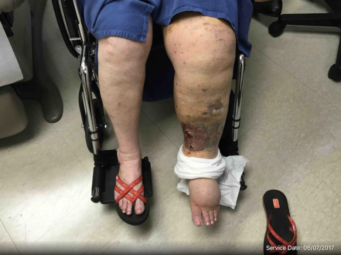
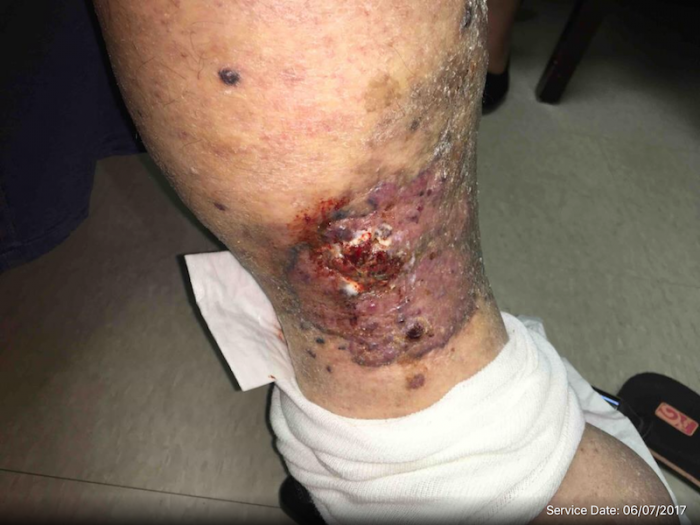
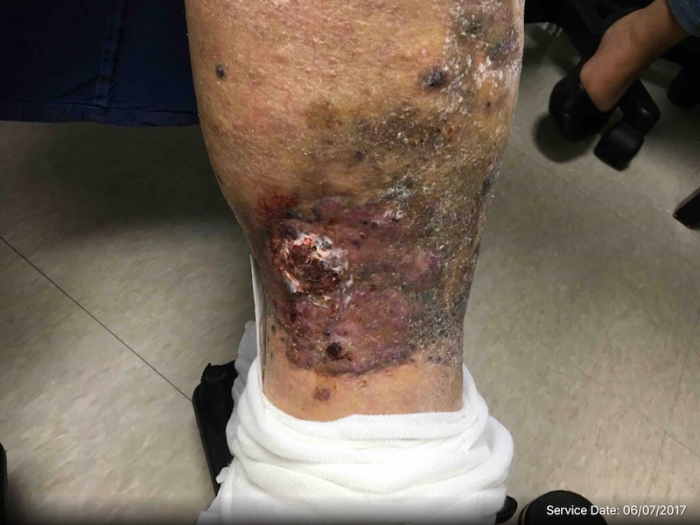
The patient had a very large fungating ulcerated violaceous lesion occupying the majority of the anterior shin. Numerous satellite lesions, mostly confluent, were present and extended circumferentially around the lower leg and proximally towards the knee. On the upper leg, diffuse petechiae were present, as well as areas of ecchymosis. No obvious satellite lesions on the upper leg. The remainder of her skin exam was normal.
LABORATORY TESTS:
Two shave biopsies were performed on the left anterior lower leg (9 mm) and left posterior lower leg (7mm).
DERMATOHISTOPATHOLOGY:
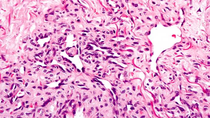
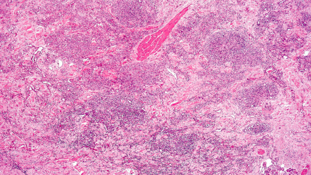
A. Left anterior lower leg: The specimen shows a haphazard proliferation of variably sized, poorly-formed vascular spaces within the superficial and mid dermis, extending diffusely to the base of the biopsy. The vascular spaces are lined by enlarged, hyperchromatic endothelial cells, some of which protrude within the lumina. Focally, there are nodular collections composed of fascicles and cords of spindled tumor cells with associated hemorrhage. CD31 and CD34 immunohistochemical stains performed with adequate controls are positive within the tumor cells. S100 and HHV-8 immunohistochemical stains are negative.
B. Left posterior lower leg: The specimen shows an ulcerated malignant vascular neoplasm composed of irregular vascular spaces lines by enlarged hyperchromatic atypical endothelial cells. Abundant associated hemorrhage is also seen. The tumor extends diffusely to the base and one lateral margin of the specimen. CD31 and CD34 immunohistochemical stains performed with adequate controls are positive within the tumor cells. S100 and HHV-8 immunohistochemical stains are negative.

DIFFERENTIAL DIAGNOSIS:
1. Chronic lymphedema/stasis dermatitis
2. Squamous cell carcinoma
3. Angiosarcoma
4. Epithelioid sarcoma
5. Ecthyma gangrenosum