CORRECT DIAGNOSIS:
Metastatic malignant melanoma with cutaneous propensity (T2aN3M1a | AJCC 2009)
DISCUSSION:
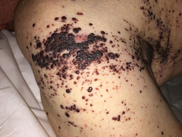
Histopathology, clinical presentation, and recent history of malignant melanoma, were diagnostic for metastatic malignant melanoma with cutaneous propensity, representing stage IV disease (T2aN3M1a | AJCC 2009). Whole-body positron emission tomography-computed tomography scan revealed no evidence of lymph node or solid organ metastasis.
Cutaneous melanoma is an aggressive disease arising from the proliferation of melanocytes. Although melanoma represents less than 5% of all skin cancers, it has a high potential for widespread metastasis and is subsequently associated with high mortality. The incidence of melanoma is growing faster than any other potentially preventable cancer in the United States with an approximately 1.9% growth annually between 2000-2009. Globally, 132,000 new cases of melanoma will arise this year with 48,000 deaths per year.
Melanoma undergoes rapid growth, causes extensive damage, and leads to high morbidity and mortality. The spontaneous eruption of extensive coalescing purple-black pigmented papulonodular lesions as a presentation of metastatic melanoma is an uncommonly reported occurrence. We have presented this case to address the need for prompt workup with a potential diagnosis of metastasis, especially in patients with a known history of cutaneous melanoma, and begin aggressive treatment to prolong survival. Physicians must maintain a high index of suspicion for cutaneous metastasis in rare eruptive presentations in patients with a previous history of malignant melanoma, as it may be the result of an underlying life-threatening event. Immunotherapy advancements over the past six years have had a profound impact on how clinicians approach metastatic melanoma treatment; we hope that this case study helps physicians in all specialties to understand the complexity of presentation for this deadly disease.
TREATMENT:
The patient was referred to oncology service for evaluation and treatment. Immunotherapy was initiated with the immune checkpoint inhibitor pembrolizumab, which targets the programmed cell death 1 (PD-1) receptor.
REFERENCES:
Bolognia, J. L., Jorizzo, J. L., & Schaffer, J. V. (2012). Dermatology (3rd ed.). Philadelphia: Elsevier Saunders.
Dasgupta, T., & Brasfield, R. (1964). Metastatic melanoma: A clinicopathological study. Cancer, 17(10), 1323-1339. https://doi.org/10.1002/1097-0142(196410)17:10<1323::AID-CNCR2820171013>3.0.CO;2-P
Fidler, I. J. (1988). The biology of melanoma metastasis. The Journal of Dermatologic Surgery and Oncology, 14(8), 875-881. https://doi.org/10.1111/j.1524-4725.1988.tb03725.x
Gerami, P., Shea, C., & Stone, M. S. (2006). Angiotropism in epidermotropic metastatic melanoma: Another clue to the diagnosis. The American Journal of Dermatopathology, 28(5), 429-433. https://doi.org/10.1097/01.dad.0000217011.24313.4b
Gupta, G. P., & Massagué, J. (2006). Cancer metastasis: Building a framework. Cell, 127(4), 679-695. https://doi.org/10.1016/j.cell.2006.11.001
Leiter, U., Meier, F., Schittek, B., & Garbe, C. (2004). The natural course of cutaneous melanoma. Journal of Surgical Oncology, 86(4), 172-178. https://doi.org/10.1002/jso.20061
Mervic, L. (2012). Time course and pattern of metastasis of cutaneous melanoma differ between men and women. PloS One, 7(3), e32955. https://doi.org/10.1371/journal.pone.0032955
Savoia, P., Fava, P., Nardo, T., Osella-Abate, S., Quaglino, P., & Bernengo, M. G. (2009). Skin metastases of malignant melanoma: A clinical and prognostic survey. Melanoma Research, 19(5), 321-326. https://doi.org/10.1097/CMR.0b013e32832d51e3
Valastyan, S., & Weinberg, R. A. (2011). Tumor metastasis: Molecular insights and evolving paradigms. Cell, 147(2), 275-292. https://doi.org/10.1016/j.cell.2011.09.024