Presenter: Ryan T. Jones, DO
Dermatology Program: MSU/Lakeland Regional Medical Center
Program Director: Mark Kuriata, D.O., F.A.O.C.D.
Submitted on: November 5, 2017
CHIEF COMPLAINT: Itchy, irritated growth on the back
CLINICAL HISTORY: The patient is a 32-year-old male who presented to our clinic with a complaint of a growth on his back which had been present for one year only. The growth is irritated and itchy but is not tender or painful. Over the course of the year the lesion has very slowly enlarged with increased itch/irritation. The patient denied any changes in color or shape since he first noticed the lesion. He also denied any other concurrent skin findings or rashes since onset. No previous treatments. He denies any previous personal or family history of skin cancer, autoimmune, or rheumatologic disease. He denies having any known medical problems other than seasonal allergies, has not had any surgeries, has no significant family history, and denies any known allergies.
PHYSICAL EXAM:
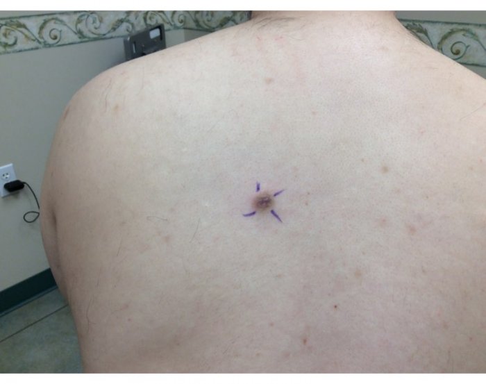
Located on the patient’s left paraspinal skin was a 1.2 x 1.0 cm well demarcated pigmented papule with overlying crust/scale, a central area of darker brown pigmentation with a surrounding lighter brown halo of pigmentation
LABORATORY TESTS:
A single 6.0mm punch biopsy was taken from the center of the lesion and was sent to the Dermatopathologist for H & E examination.
DERMATOHISTOPATHOLOGY:
Histologic examination reveavled hyperkeratosis, epidermal acanthosis, basal epidermal hyperpigmentation and a dense dermal proliferation of fibrohistiocytic cells. The fibrohistiocytic cells had a curlicue growth pattern with peripheral collagen trapping. No cytologic atypia was noted. Immunohistochemical staining was performed (results withheld and will be discussed at end of the case presentation).
DIFFERENTIAL DIAGNOSIS:
1. Atypical/Dysplastic Nevus
2. Malignant Melanoma
3. Irritated/Inflamed Seborrheic Keratosis
4. Dermatofibroma
5. Inflamed Epidermal Inclusion Cyst