CORRECT DIAGNOSIS:
Cutaneous Piloleiomyomas consistent with Reed’s Syndrome
DISCUSSION:
Reed’s syndrome is an autosomal dominant genodermatosis characterized by multiple cutaneous leiomyomas and uterine leiomyomas (also known as multiple cutaneous and uterine leiomyomatosis [MCUL]), which is associated with an increased risk of an aggressive type II papillary renal cell carcinoma developing in a subset of affected individuals. Familial studies have revealed predisposition to Reed’s syndrome due to loss of heterozygosity mutations of the fumarate hydratase gene, localized to a 14-cM region of chromosome 1q42.3-43. Fumarate hydratase encodes the enzyme fumarase involved in the Krebs cycle, as it converts fumarate to malate and is speculated to function as a tumor suppressor gene. Tumorigenesis is thought to occur due to loss of function mutations of fumarate hydratase resulting in metabolic dysfunction and subsequent hypoxia, which in turn promotes cellular transformation and tumor formation. Reed’s syndrome affects males and females equally, however, the condition may be recognized more frequently in women due to the symptomatology of uterine fibroids.
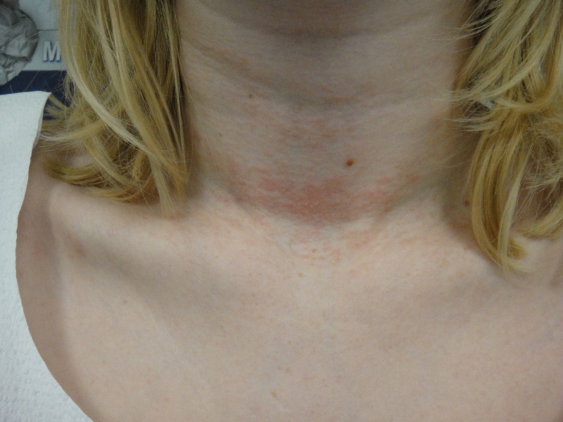
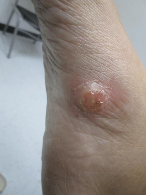
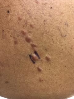
Clinically, patients with Reed’s syndrome can present with leiomyomas of the uterus and skin. Uterine leiomyomas tend to be larger in size, more numerous, and present earlier in affected women in comparison to the general population. Menorrhagia and pelvic pain often result in women undergoing hysterectomy or myomectomy at a younger age. Cutaneous leiomyomas, the most common finding, are benign tumors arising from the arrector pili muscle of hair follicles (piloleiomyomas), vascular smooth muscle (angioleiomyomas), and tunica dartos muscle of the scrotum or mammary muscles of the nipples (genital leiomyomas). Leiomyomas appear as small, solitary or multiple, firm, skin-colored to pink-brown, smooth-surfaced papules or nodules, frequently associated with pain that can be exacerbated by exposure to cold or pressure. Lesions are commonly located to the extensor surface of the extremities and trunk, and may be distributed bilaterally or in a linear, zosteriform or dermatomal fashion. Biopsy of lesions should be performed as histopathologic examination is required to confirm the diagnosis of cutaneous leiomyomas. Characteristic histologic findings include smooth muscle fibers with eosinophilic cytoplasm and blunt ended, cigar-shaped nuclei arranged in fascicles and interspersed between collagen within the dermis (desmin and actin positive). Proposed criteria for diagnosis of Reed’s syndrome include: multiple cutaneous leiomyomas with at least one histologically confirmed lesion or a single leiomyoma in the presence of a positive family history of MCUL.
A high index of suspicion for Reed’s syndrome is essential in those patients presenting with multiple cutaneous leiomyomas to ensure patients are appropriately evaluated/regularly screened for occult renal cell carcinoma and uterine disease (pelvic/renal ultrasound, urinalysis). Early detection and treatment of renal cell malignancy can significantly improve patient survival. Other screening recommendations include but are not limited to complete physical examination with pelvic examination and detailed history with emphasis on family and surgical history. It is also recommended that patients have molecular genetic testing with sequencing analysis of the FH gene. Genetic counseling should be provided to affected individuals and their family members.
TREATMENT:
Treatment of cutaneous leiomyomas is indicated for symptomatic or numerous lesions, which may be distressing to patients. Pain may be managed by medications such as oral nifedipine or phenoxybenzamine, and botulinum toxin A, which function to inhibit smooth muscle contraction and subsequent nerve stimulation. Medications such as gabapentin may also be considered in those patients presenting with painful lesions. Complete removal of lesions with surgical excision is considered the gold standard in individuals presenting with few cutaneous leiomyomas. Other effective treatment modalities include carbon dioxide laser ablation. A multidisciplinary approach is highly recommended with coordination of care between the dermatologist, internist, gynecologist, as well as nephrologist/oncologist if renal cell tumor is detected. Renal cell carcinomas require total nephrectomy as the papillary subtype is quite aggressive and has the potential to metastasize quickly.
We recommended our patient to follow-up with her primary care physician/internist for complete physical examination and laboratory evaluation including CBC, CMP, and urinalysis. We also ordered abdominal/pelvic CT imaging to screen for possible renal cell tumor.
REFERENCES:
Emer JJ, Solomon S, Mercer SE. Reed’s syndrome: a case of multiple cutaneous and uterine leiomyomas. J. Clin. Aesthet. Dermatol. 2011 Dec;4(12):37-42.
Kontochristopoulos G, Kouris A, Balamoti E, et al. A case of Reed’s syndrome: an underdiagnosed tumor disorder. Case Rep Dermatol. 2014;6(2):189-93.
Alam NA, Barcaly E, Rowman AJ, et al. Clinical features of multiple cutaneous and uterine leiomyomatosis: an underdiagnosed syndrome. Arch of Derm. 2005;141:199-206.
Christenson LJ, Smith K, Arpey CJ. Treatment of multiple cutaneous leiomyomas with CO2 Laser Ablation. Dermatol Surgery. 2000; 26(4):319-22.
Holst VA, Junkins-Hopkins JM, Elenitsas R. Cutaneous smooth muscle neoplasms: Clinical histologic findings, and treatment options. J Am Acad Dermatol, 2002;46:477-90.