CORRECT DIAGNOSIS:
Cutaneous Leishmaniasis
DISCUSSION:
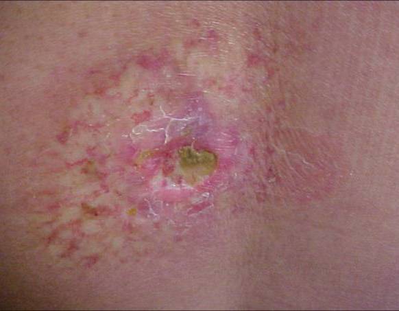
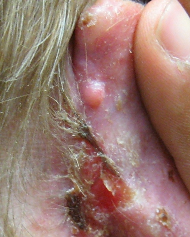
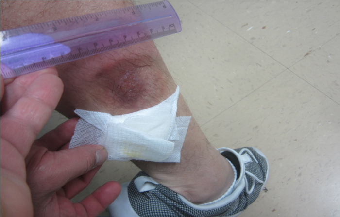
Leishmaniasis is a disease caused by an intracellular parasite that causes multiple clinical symptoms. According to the World Health Organization, it is one of the seven most important tropical zoonoses. Its manifestations vary in degree of severity depending on the species involved and the immune response from patients. These can present from self-limited skin ulcers to the most severe form where infection can cause multisystem failure secondary to hemorrhage and thrombocytopenia. Classification based on clinical symptoms includes: cutaneous, mucocutaneous and visceral. Our patient presented with the cutaneous variant.
Cutaneous Leishmaniasis can be caused by L. tropica in the Eastern Hemisphere or L. Mexicana in Mexico and Central America. Insect bites usually occur in exposed areas most commonly on ears, nose, upper extremities and ankles. The parasite has an incubation period of 1-4 weeks but may last several years. Initially, it presents with erythematous papules with pruritus in some patients. The lesion size can range from 1 to 10 millimeters. After 48 hours, it converts to a vesicle then into a pustule. Eventually, it may break due to trauma, revealing an ulcer with round borders and sharp edges. These ulcers can last from months to several years.
Diagnosis is usually made by clinical symptoms and epidemiological context. The protozoa can be found on skin scrapings of lesions. Histopathological studies reveal epidermal hyperplasia or atrophy with infiltration of macrophages, lymphocytes and plasma cells with localized necrotic areas. Parasites known as amastigotes, can be found intracellularly within cytoplasmic vacuoles on histiocytes during early stages. During late stages, a lympho-histiocytic infiltrate is seen within infected macrophages. Although PCR is not available in many countries where Leishmania is endemic, it shows 100% specificity for CL.
TREATMENT:
The approach to management of cutaneous leishmaniasis (CL) begins with establishing the clinical severity of infection.
Uncomplicated versus complicated infection:
- No mucosal involvement
- Single lesion or a few lesions
- Small lesion size (eg, <1 cm)
- Immunocompetent host
Whom to treat:
Clinical observation (in the absence of treatment) is reasonable for immunocompetent patients with uncomplicated lesions that are healing spontaneously whose infection is either known to be caused by a Leishmania species not associated with increased risk for ML.
Local therapy is reasonable for patients with uncomplicated CL who are not already healing spontaneously and/or who would like to pursue therapeutic intervention. Local therapy is appropriate for management of Old World CL as well as for treatment of New World CL caused by species unlikely to cause disseminated infection. Topical Paromycin or intralesional pentavalent antimonial can be used.
Systemic therapy is warranted for patients with complicated CL, for immunocompromised patients, for patients with spontaneously healing or recently healed lesions whose infection is known to be caused by a Leishmania species associated with increased risk for ML.
REFERENCES:
Torres-Guerrero E, Quintanilla-Cedillo MR, Ruiz-Esmenjaud J, Arenas R. Leishmaniasis: a review. F1000Research. 2017;6:750. doi:10.12688/f1000research.11120.1.
Vargas-Martínez F, Torres-Guerrero E, Quintanilla-Cedillo MR, et al. : Leishmaniasis en México. Academia Mexicana de Dermatología, Colegio de Dermatólogos de Yucatán A. C., Fundación Mexicana para la Dermatología, Universidad Autónoma de Campeche y Secretaría de Salud, México.2013.
Andrade-Narváez FJ, Vargas-González A, Canto-Lara SB, et al. : Clinical picture of cutaneous leishmaniases due to Leishmania (Leishmania) mexicana in the Yucatan peninsula, Mexico. Mem Inst Oswaldo Cruz. 2001;96(2):163–7. 10.1590/S0074-02762001000200005
Incháustegui A, et al. De la leishmaniosis americana y de la úlcera de los chicleros en México. Tesis, Universidad Nacional de México.1918.
Vera-Izaguirre D, Vega-Memije E, Quintanilla Cedillo M, et al. : Leishmaniasis revisión.2006;4(4):252–260.