Presenter: Jeffrey Harbold, DO, Carlos A. Rivera MD, Rick Lin, DO, Michael Hohnadel, DO, Thomas L Davis, MD
Dermatology Program: South Texas Dermatology Residency Program, Bay Area Corpus Christi Medical Center
Program Director: Rick Lin, DO MPH
Submitted on: October 7, 2019
CHIEF COMPLAINT: ¨I have skin lesions on both arms”
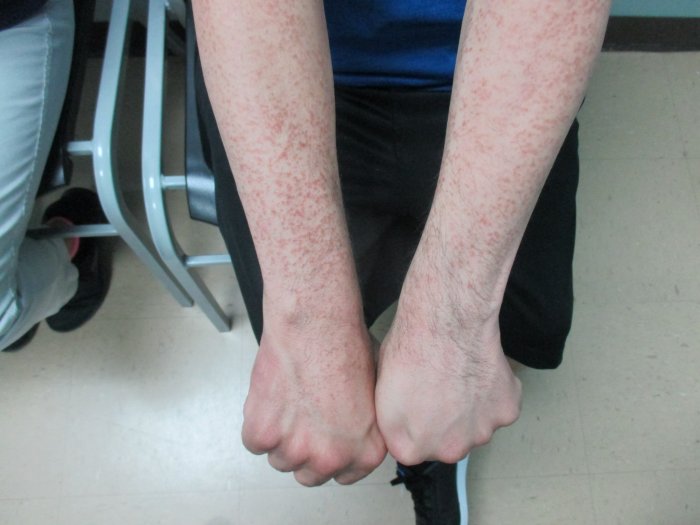
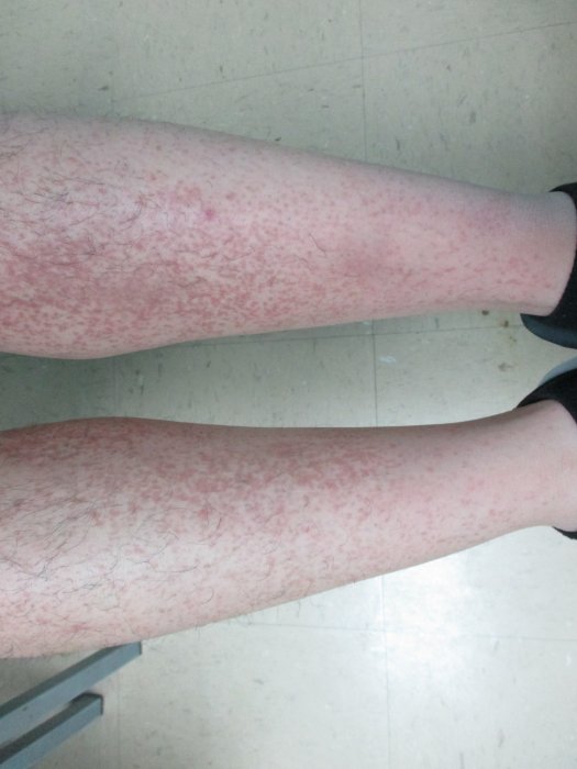
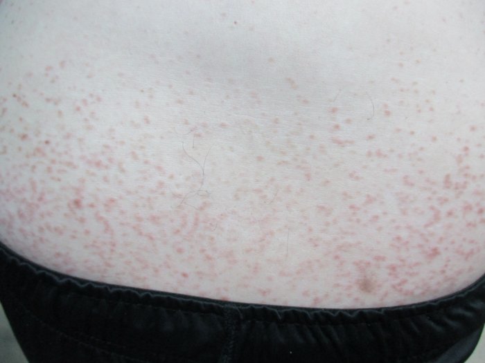
CLINICAL HISTORY: A 45-year-old Hispanic male with a past medical history of Down syndrome was referred to our clinic with an 8-year history of numerous crops of hyperpigmented confluent smooth papules. Lesions originated on the flexural surfaces of both arms with subsequent spread to the lower torso and legs, with facial sparing. The patient reported occasional mild pruritus but denied any associated pain. Other dermatologic history include biopsy-confirmed scabies in 2015 successfully treated with topical permethrin and a history of rosacea controlled with metronidazole gel and an occasional oral minocycline. There was no significant dermatologic family history reported.
PHYSICAL EXAM:
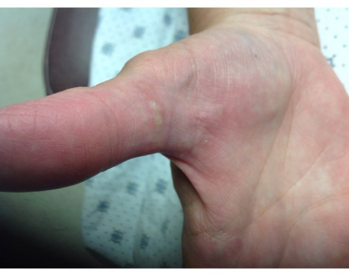
Complete physical examination revealed diffuse pink, flat topped papules measuring 2 to 3 mm in diameter. Lesions were symmetrically distributed on the flexor arms, upper anterolateral legs, lower anterior legs and lower torso. The patient also demonstrated physical exam findings consistent with Down syndrome, including; epicanthal fold, short phalanges and flat-bridged nose. Genetic testing performed in infancy was positive for trisomy 21.

LABORATORY TESTS: N/A
DERMATOHISTOPATHOLOGY:
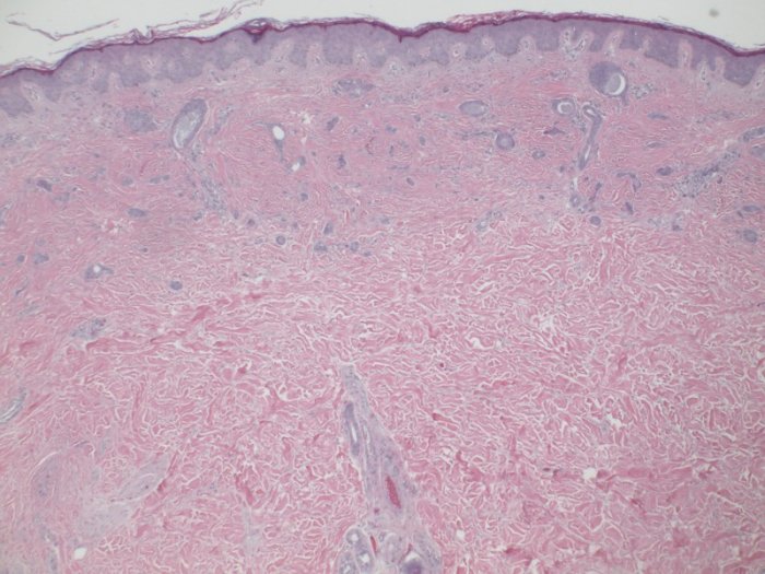
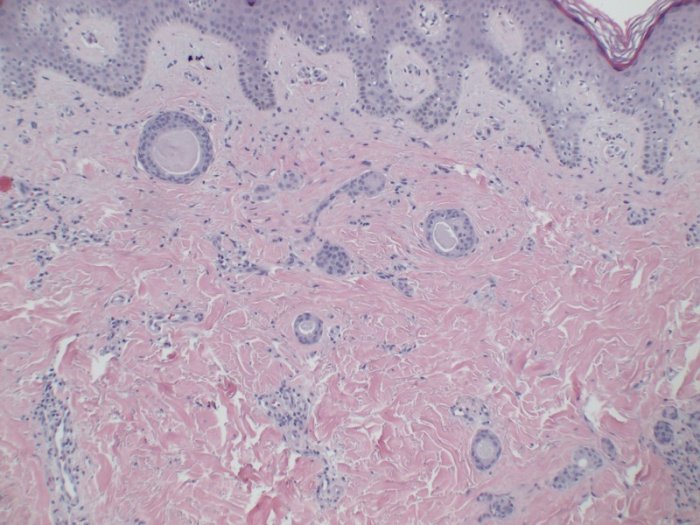
A 4mm biopsy was obtained from the right hip and microscopic examination demonstrated multiple duct-like structures lined by flat epithelial cells in a fibrous stroma. Low view-microscopy demonstrating fibrous stroma, multiple duct-like structures with a tadpole appearance lined by multiple layers of cells. High view-microscopy demonstrating fibrous stroma, multiple duct-like structures with a tadpole appearance lined by multiple layers of cells.
DIFFERENTIAL DIAGNOSIS:
1. Lichen Planus
2. Cowden disease
3. Angiofibroma
4. Fibrofolliculoma
5.Eruptive syringoma
6. Trichoepithelioma