Presenter: Soham Chaudhari, DO, Carlos Rivera, MD, Thomas L Davis, MD, Rick Lin, DO
Dermatology Program: South Texas Dermatology Residency Program
Program Director: Rick Lin, DO
Submitted on: December 1, 2018
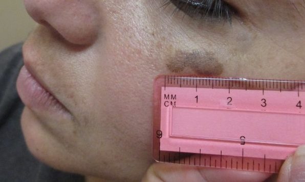
CHIEF COMPLAINT: Brown solitary macule on the left cheek
CLINICAL HISTORY: A 38-year-old female with no past medical history who was referred to our dermatology clinic from her PCP for a lesion that has been present for 5 years. The patient denies any surgical history, medication use, allergies, or any type of family history of skin cancer. The patient also denies smoking, drinking alcohol, or use of illicit drugs.
PHYSICAL EXAM:
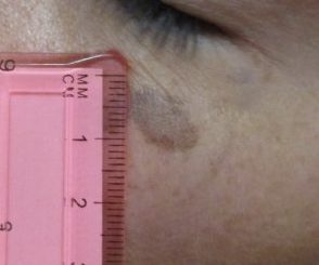
Physical examination revealed a solitary melanocytic macule on left zygoma measuring 1.8 cm x 0.6 cm, with a dark border on the inferior portion of the left side and a reticular pigment network on the right side. The patient had ephelides around the lesion and in malar region.

DERMATOHISTOPATHOLOGY:
Histopathologic studies revealed epidermis mildly papillated and pigmented with a lymphocytic infiltrate. Also revealed was a proliferation of atypical melanocytes along the epidermis arranged as focally confluent solitary units along an effaced dermoepidermal junction.
DIFFERENTIAL DIAGNOSIS:
1. Seborrheic keratosis.
2. Melanocytic nevus.
3. Solar lentigo.
4. Melanoma in-situ.