CORRECT DIAGNOSIS:
Melanoma in-situ.
DISCUSSION:
A seborrheic keratosis usually presents as small, hyperpigmented scaling lesions. Its size ranges in millimeters and may appear as single or multiple lesions.4 These lesions are usually recognized visually and not often biopsied. It is not uncommon for these lesions to appear on the face, extremities or trunk. Clinical attention is usually given when patients present with symptoms such as inflammation, or itchiness.5 No treatment is needed except for aesthetic or symptomatic reasons. Shave biopsy, electrodesiccation and curettage, or cryotherapy are some of the treatment options for removing seborrheic keratosis.
A retrospective study elucidated the use of dermoscopy when excluding melanoma in patients with suspected seborrheic keratosis. Clues that support the diagnosis of melanoma included a pigment network, pseudopods, the blue-black sign and/or blue/white veil.6 It is important to note that even with the use of dermoscopy 27% of patients from this study were incorrectly diagnosed.6
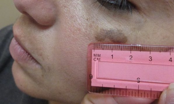
We presented a case of a patient with an initial clinical picture of seborrheic keratosis; although upon closer inspection with deep section histological analysis, the diagnosis of early melanoma in-situ was supported. Therefore, it is important for clinicians to perform a thorough evaluation that will provide the correct management for our patients.
TREATMENT:
The patient elected to have the melanoma in-situ excised by a plastic surgeon.
CONCLUSION:
The case we presented highlights the importance of being cognizant about the alternative diagnosis when patients present with a clinical picture of seborrheic keratosis. Melanoma should be included as a differential diagnosis and excluded from a histopathologic study. Physicians should be aware that melanoma can be disguised as another condition and a thorough workup should not be overlooked in order to avoid catastrophic consequences for these patients. Increased awareness of this occurrence may help physicians in the early recognition and accurate treatment of seborrheic keratosis-like melanoma.
REFERENCES:
Argenziano, Giuseppe. Melanoma Simulating Seborrheic Keratosis: A Major Dermoscopy Pitfall. Archives of dermatology. March 2003. Volume: 139 Issue: 3 Page: 389-391. PMID: 12622648.
Tiffany Y. Chen, Annie O. Morrison, Clay J. Cockerell. Cutaneous malignancies simulating seborrheic keratoses: An underappreciated phenomenon? Journal of Cutaneous Pathology. 2017 Sep;44(9):747-748. PMID: 28589622
Thomas I, Kihiczak NI, Rothenberg J, Ahmed S, Schwartz RA. Melanoma within the seborrheic keratosis. Dermatology Surgery.2004 Apr;30(4 Pt 1):559-61.
Karadag, A.S., Parish, L.C. The status of seborrheic keratosis. Clinics in Dermatology. Volume 36, Issue 2, March – April 2018, Pages 275-277.
Leonid Izikson, BS; Arthur J. Sober, MD; Martin C. Mihm Jr, MD, FRCP; et al. Prevalence of Melanoma Clinically Resembling Seborrheic Keratosis. Arch Dermatol. 2002;138(12):1562-1566. PMID:12472343
CARRERA, C; et al. Dermoscopic Clues for Diagnosing Melanomas That Resemble Seborrheic Keratosis. JAMA Dermatology. United States, 153, 6, 544-551, June 1, 2017. ISSN: 2168-6084. PMID: 28355453