Presenter: Eric Sandrock, DO, Thomas L Davis, MD, Rick Lin, DO
Dermatology Program: South Texas Dermatology Residency, HCA Healthcare Corpus Christi Medical Center – Bay Area Program
Program Director: Rick Lin, DO MPH FAOCD
Submitted on: September 20, 2023
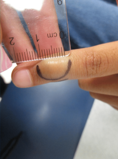
CHIEF COMPLAINT: “I have a hard lump on my finger “
CLINICAL HISTORY: A 10-year-old male with no significant past medical or surgical history presents to our clinic for a solitary lesion overlying the distal left index finger. He describes it as “hard” and stated it has been present and stable for the past year. He denied any trauma to the area, restriction in range of motion, or tenderness to palpation. He denied any recent weight loss, fevers, or chills. He denied any personal or family history of similar lesions or skin cancer.
PHYSICAL EXAM:
Physical examination showed a solitary, 1 cm at longest dimension, firm, fixed, and smooth nodule on the distal interphalangeal joint of the left index finger. No erythema or tenderness on palpation was noted.

LABORATORY TESTS: N/A
DERMATOHISTOPATHOLOGY:
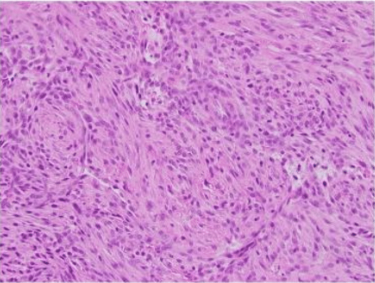
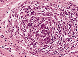
A 3 mm punch biopsy of the lesion was performed. Histopathologic examination revealed nodular collections of histiocytes in the reticular dermis, extending to the specimen’s edges. Many of the histiocytes displayed foamy cytoplasm within a fibrotic stroma, with a rare multinucleated histiocyte present. No mitotic figures were identified. Immunostains for S100, EMA, and CK8/18 were negative, and the Fite stain was also negative.
DIFFERENTIAL DIAGNOSIS:
1. Digital Mucous Cyst
2. Callus
3. Calcinosis Cutis
4. Rheumatoid Nodule
5. Juvenile Idiopathic Arthritis
6. Digital Papillary Adenocarcinoma
7. Gouty Tophus
8. Digital Fibromatosis
9. Deep Fungal Infection
10. Atypical Mycobacterial Infection
10. Foreign Body