Presenter: Paul Vance, DO, Thomas L Davis, MD, Michael Hohnadel, DO
Dermatology Program: South Texas Dermatology Residency, HCA Healthcare Corpus Christi Medical Center – Bay Area Program
Program Director: Rick Lin, DO MPH FAOCD
Submitted on: September 24, 2023
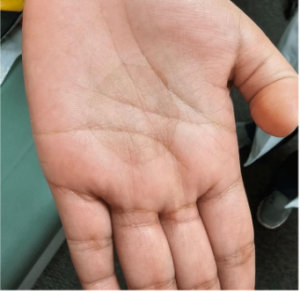
CHIEF COMPLAINT: “I have a spot growing in size on my left hand”
CLINICAL HISTORY: An 11-year-old Hispanic female presented to our clinic for evaluation of a growing dark spot on the palm of her left hand. The patient states that the lesion has been present for the past several months and seems to be getting bigger. The patient endorses occasional itchiness, but no pain. No one else in the household had similar lesions. The patient denies any surgical history, medication use, allergies, recent travel, or personal or family history of skin cancer.
PHYSICAL EXAM:
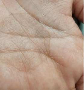
Physical examination revealed a solitary, well-defined 3 cm x 3 cm hyperpigmented patch on the palmar surface of the patient’s left hand with minimal scale. There is also a noticeably darker border.
LABORATORY TESTS: N/A
DERMATOHISTOPATHOLOGY:
A shave biopsy was performed to obtain a sample of the pigmented area. Histopathological studies revealed numerous short, segmented hyphae and spores in the superficial stratum corneum. The epidermis and dermis are largely unremarkable.
DIFFERENTIAL DIAGNOSIS:
1. Acral Lentiginous Melanoma
2. Junctional nevus
3. Post Inflammatory Hyperpigmentation
4. Tinea Infection
5. Stain