CORRECT DIAGNOSIS:
Tinea Nigra
DISCUSSION:
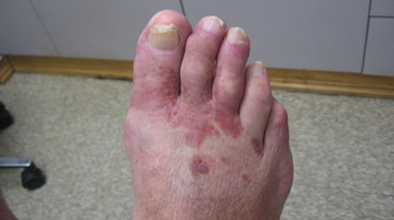
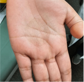
Superficial fungal infections of the hair, skin, and nails are significant contributors to global morbidity. Among these, tinea nigra stands out as an uncommon mold infection affecting the skin of the palms and/or soles. Characterized by dark patches – black or brown in color – this condition predominantly occurs in tropical regions and is more likely to affect individuals with excessive sweating.
Tinea nigra is primarily caused by molds, including Phaeoannellomyceas, Hortaea, or Exophiala werneckii, which are dematiaceous fungus typically found in soil. The infection occurs when these fungi penetrate the skin, leading to localized pigmentation changes. Clinically, tinea nigra manifests as asymptomatic, pigmented patches on the palms or soles. Dermoscopy can be particularly useful for diagnosis, revealing fine, light brown strands or “pigmented spicules” arranged in a reticular-like pattern with uniform brown coloration. This presentation is distinct from acral melanomas and melanocytic nevi, which show a parallel ridge and furrow pattern on dermoscopy.
Diagnosis of tinea nigra often involves superficial skin scraping for microscopic examination with potassium hydroxide or a biopsy. Microscopy typically reveals pigmented hyphae, leading to the diagnosis of tinea nigra. Histological examination shows numerous short, segmented hyphae and spores in the superficial stratum corneum, often appearing brown-yellow color on routine hematoxylin and eosin stains. Importantly, the epidermis and dermis remain largely unremarkable, distinguishing it from other skin conditions. The treatment of tinea nigra generally includes the application of topical antifungal agents for two to four weeks.
Prior reports have highlighted cases of tinea nigra in patients with travel histories to tropical areas. For instance, a 13-year-old presented with a brown patch following a trip to the Greek islands, ultimately diagnosed with tinea nigra. In our clinical experience, we encountered a patient whose initial clinical presentation mimicked a melanocytic superficial lesion. A biopsy confirmed the diagnosis of tinea nigra, and the condition resolved with consistent use of a topical antifungal cream.
TREATMENT:
The classic treatment of tinea nigra involves topical antifungal cream applied till resolution is achieved, typically in 2-4 weeks. Our patient was treated with topical ketoconazole 2% cream applied twice daily till clearance was reached at around 4 weeks.
CONCLUSION:
Tinea nigra, while relatively uncommon, presents significant diagnostic challenges due to its clinical resemblance to more serious conditions, such as acral melanomas. This case underscores the necessity for clinicians to include tinea nigra in the differential diagnosis when encountering pigmented patches on acral surfaces.
Its dark pigmentation can easily lead to misinterpretation as a melanocytic lesion, highlighting the importance of thorough evaluation. Dermoscopy is a valuable tool that greatly enhances diagnostic accuracy, allowing physicians to identify the unique features of tinea nigra—namely, its reticular pattern and uniform pigmentation.
Moreover, it is crucial for healthcare providers to conduct a comprehensive workup to rule out neoplastic causes, including melanoma-like lesions and black eschars. Failure to do so could result in detrimental consequences for patients. As global travel increases, the recognition of tinea nigra and similar infections in patients with recent exposure to tropical environments becomes ever more vital. By maintaining a high index of suspicion and utilizing appropriate diagnostic techniques, healthcare professionals can ensure timely and effective treatment for this fungal infection.
REFERENCES:
Belmokhtar, Z., Djaroud, S., Matmour, D., & Merad, Y. Atypical and unpredictable superficial mycosis presentations: A narrative review. Journal of Fungi. 2024 Apr; 10(4):295. [PMID 38667966]
Martínez-Ortega, JI., Fernández-Reyna, I., Atoche Dieguez, CE., Espinosa Alonzo, L., & Ramirez Cibrian, AG. Tinea nigra: Clinical and diagnostic guidance. Cureus. 2024 Aug; 16(8). [PMID 39246895]
Nazzaro, G., Ponziani, A., & Cavicchini, S. Tinea nigra: A diagnostic pitfall. Journal of the American Academy of Dermatology. 2016 Dec; 75(6). [PMID 27846967]