CORRECT DIAGNOSIS:
Lichen Planus Pigmentosus-Inversus
DISCUSSION:
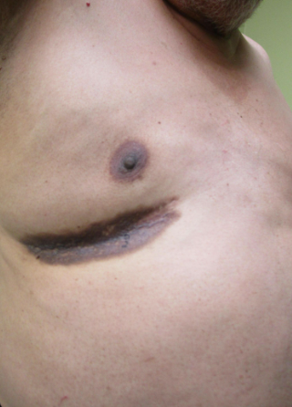
Lichen Planus Pigmentosus Inversus (LPPI) is an uncommon variant of lichen planus that primarily affects darker-skinned individuals (skin types III and IV), particularly middle-aged Indian and Middle Eastern females. While Lichen Planus Pigmentosus (LPP) typically presents as brown to violaceous or gray-brown macules, patches, or plaques in sun-exposed areas. LPPI is characterized by lesions that are limited to intertriginous areas such as the axilla, groin, and inframammary regions. The first description of this unusual variant occurred in 2001 by Pock, highlighting its occurrence in Caucasians from Central Europe. LPPI can manifest at any age but has been reported in individuals ranging from 15 to 82 years old.
LPP usually presents asymptomatically, though some patients may experience mild pruritus or a burning sensation. The lesions can evolve from macules to diffuse or reticulate pigmentation and typically do not involve hair, nails, mucosal surfaces, or palmoplantar areas. The course is chronic and progressive, with remissions and exacerbations, and can last from six months to three years. LPPI is associated with various conditions, including hepatitis C, diabetes mellitus, autoimmune disorders (such as hypothyroidism and vitiligo), hyperlipidemia, chronic urticaria, and tuberculosis.
The pathogenesis of LPPI is thought to involve T lymphocyte-mediated cytotoxic activity against basal keratinocytes, with potential triggers including external mechanical stimuli (e.g., friction from tight clothing) and the use of certain topical products (such as mustard oil and henna). Recent reports have also suggested associations with radiotherapy, paraneoplastic syndromes, and COVID-19 vaccination.
Diagnosis of LPPI is primarily clinical, based on the characteristic appearance of the lesions and patient history. A thorough evaluation for associated conditions is also critical. A skin biopsy may be performed to confirm the diagnosis and rule out other dermatological conditions. Histopathological examination typically reveals key features such as a band-like infiltrate of lymphocytes in the upper dermis, vacuolar degeneration at the epidermal-dermal junction, and pigmentary incontinence due to melanin leakage, supporting a diagnosis of LPPI.
In this case, our patient’s lesions, which developed after a hospitalization for a urinary tract infection complicated by sepsis, reflect the common trigger of external mechanical stimuli in LPPI, especially given their location in intertriginous areas. Given the associations of LPPI with systemic conditions like diabetes and autoimmune disorders, the patient’s medical history raises the possibility of a multifactorial etiology. Therefore, a comprehensive clinical evaluation remains essential for managing his skin condition and monitoring for potential complications related to underlying systemic diseases.
TREATMENT:
The management of Lichen Planus Pigmentosus Inversus (LPPI) primarily focuses on alleviating symptoms and minimizing the appearance of lesions. Topical corticosteroids, particularly high-potency formulations, are often the first line of treatment, effectively reducing inflammation and pruritus associated with the condition. In cases where corticosteroids are not preferred or for sensitive areas, calcineurin inhibitors such as tacrolimus or pimecrolimus may be utilized as alternatives. For patients with widespread lesions, phototherapy, specifically narrowband ultraviolet B (NB-UVB) therapy, can be beneficial in improving pigmentation and reducing inflammation. In instances of extensive or resistant LPPI, systemic treatments, including corticosteroids or immunosuppressive agents like azathioprine or methotrexate, may be considered. Additionally, educating patients about avoiding potential triggers—such as friction from tight clothing or specific topical products—can aid in managing and preventing exacerbations. Regular monitoring through follow-up appointments is crucial to assess the progression of lesions and the effectiveness of the treatment plan.
For our patient, a tailored approach was implemented that incorporated topical corticosteroids to address pruritus and inflammation, alongside careful monitoring of any underlying conditions. Shared decision-making led to the establishment of goals: to decrease pigmentation, improve aesthetic appearance, and enhance quality of life by reducing emotional stress. The patient was initially started on triamcinolone acetonide 0.025% ointment twice daily for four weeks; however, he did not experience noticeable improvement. Consequently, he was switched to hydroquinone 4% for nightly application, but he discontinued it due to worsening lesions and a burning sensation. Ultimately, the patient was prescribed tacrolimus 0.1% ointment to be applied twice daily, which resulted in noticeable improvement in hyperpigmentation after four weeks of consistent use.
CONCLUSION:
LPPI is a distinctive variant of lichen planus that primarily affects darker-skinned individuals, particularly middle-aged Indian and Middle Eastern females. Clinically, it presents as asymptomatic brown to violaceous macules and plaques localized to intertriginous areas, such as the axilla, groin, and inframammary folds, sparing sun-exposed skin. The association with various systemic conditions, including hepatitis C and autoimmune disorders, underscores the importance of a comprehensive clinical evaluation.
The chronic and progressive nature of LPPI, characterized by remissions and exacerbations, complicates its management and necessitates careful monitoring. Understanding the potential triggers—such as friction, certain topical agents, and recent reports linking LPPI to external stimuli—can aid in patient education and management. Overall, LPPI represents a unique clinical entity that requires a nuanced approach to diagnosis and treatment, with ongoing research necessary to elucidate its underlying pathophysiology and optimal management strategies.
REFERENCES:
Barros, H. R., Almeida, J. R., Mattos e Dinato, S. L., Sementilli, A., & Romiti, R. (2013). Lichen planus pigmentosus inversus. Anais Brasileiros de Dermatologia, 88(6 Suppl 1), 146-149. https://doi.org/10.1590/abd1806-4841.20132082 [PMID: 24474191]
Bolognia JL, Schaffer JV, Cerroni L. (2018). Dermatology. Elselvier.
Cohen, P. R., Erickson, C. P., & Calame, A. (2024). Lichen Planus Pigmentosus Inversus: A Case Report of a Man Presenting With a Pigmented Lichenoid Axillary Inverse Dermatosis (PLAID). Cureus, 16(3), e56995. https://doi.org/10.7759/cureus.56995 [PMID: 37198852]
Edek, Y. C., Tamer, F., & Öğüt, B. (2022). Lichen planus pigmentosus inversus with nail involvement following COVID-19 vaccination: A case report. Dermatology and Therapy, 35(11), e15809. https://doi.org/10.1111/dth.15809 [PMID: 36115338]
Robles-Méndez, J. C., Rizo-Frías, P., Herz-Ruelas, M. E., Pandya, A. G., & Ocampo Candiani, J. (2018). Lichen planus pigmentosus and its variants: review and update. International Journal of Dermatology, 57(5), 505-514. https://doi.org/10.1111/ijd.13925 [PMID: 29288998]
Khopkar, U., & Choudhary, S. (2018). Lichen Planus Pigmentosus: An Overview. Indian Journal of Dermatology, Venereology and Leprology, 84(1), 14-23. https://doi.org/10.4103/ijdvl.IJDVL_300_16 [PMID: 29396516]
Pock, L. (2001). Lichen Planus Pigmentosus Inversus: A New Variant of Lichen Planus. Archives of Dermatology, 137(10), 1354-1358. https://doi.org/10.1001/archderm.137.10.1354 [PMID: 11529490]
Ezzedine, K., & Watt, T. (2014). Lichen Planus and its Variants. Dermatologic Clinics, 32(3), 351-360. https://doi.org/10.1016/j.det.2014.03.008 [PMID: 25065902]
Ranjan, K., & Kumar, S. (2017). Topical Therapy for Lichen Planus: A Review. The Clinical Review of Dermatology, 19(2), 138-146. https://doi.org/10.1016/j.clindermatol.2017.01.001 [PMID: 28153480]
Morelli, J. G., & Kogan, R. (2019). Lichen Planus: A Comprehensive Review. The Journal of Clinical and Aesthetic Dermatology, 12(5), 32-38. https://www.ncbi.nlm.nih.gov/pmc/articles/PMC6503151/ [PMID: 31030062]


