Presenter: Eric Sandrock, DO, Thomas L Davis, MD, Rick Lin, DO
Dermatology Program: South Texas Dermatology Residency, HCA Healthcare Corpus Christi Medical Center – Bay Area Program
Program Director: Rick Lin, DO MPH FAOCD
Submitted on: September 18, 2024
CHIEF COMPLAINT: “A new pimple grew on my arm “
CLINICAL HISTORY: A 67-year-old male with a medical history of hypertension, coronary artery disease, non-melanoma skin cancers, lower esophageal adenocarcinoma, and intracranial arteriovenous malformations, presented to our clinic to discuss biopsy results from a lesion on his left cheek. The biopsy confirmed the presence of a nodular and pigmented basal cell carcinoma. During this visit, the patient also reported a new papule on his left proximal forearm, which he indicated had been present for only a few weeks. He denied any bleeding or discomfort associated with the new lesion.
The patient’s surgical history includes a coronary artery bypass graft (CABG) procedure involving four vessels, as well as previous excisions for skin cancers: a basal cell carcinoma on the right shoulder that underwent Mohs micrographic surgery and a squamous cell carcinoma on the left forearm that also received Mohs treatment. His current medication regimen includes Nexium and Hydrocortisone, and he denied any allergies to medications. The patient reports no pertinent family history. He denies smoking, illicit drug use, or alcohol consumption.
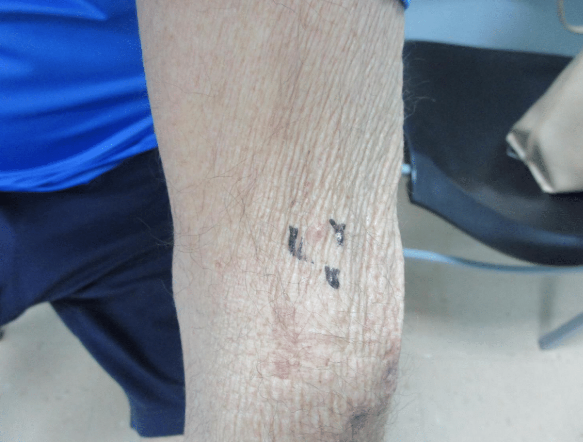
PHYSICAL EXAM:
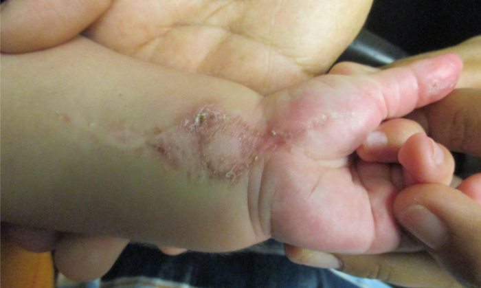
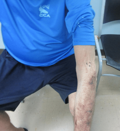
Physical examination demonstrated a skin-colored 2mm papule on a slightly erythematous base on the left proximal forearm.
LABORATORY TESTS: N/A
DERMATOHISTOPATHOLOGY:
A shave biopsy revealed dermal infiltration by nests and small cords of enlarged polygonal hyperchromatic cells. Tumor nests contained multiple small luminal formations, with focal necrosis and noted mitotic activity. Importantly, there was no connection to the overlying epidermis.
Immunohistochemical staining showed positive results for carcinoembryonic antigen (CEA), BerEP4, cytokeratin AE1/A3, CDX-2, and cytokeratin 20 (CK20). The tumor was negative for p63, CK7, synaptophysin, GATA-3, and chromogranin.
DIFFERENTIAL DIAGNOSIS:
-
- Milia
- Hemangioma
- Basal Cell Carcinoma
- Arthropod Bite
- Papular Mucinosis
- Lichen Nitidus