CORRECT DIAGNOSIS:
Metastatic Adenocarcinoma
DISCUSSION:
The diagnosis of metastatic adenocarcinoma is supported by the histopathologic and immunohistochemical findings from the shave biopsy. The presence of nests of polygonal hyperchromatic cells, luminal formations, and focal necrosis suggests an aggressive tumor. The IHC markers play a crucial role in determining the primary site of the adenocarcinoma.
Thyroid Transcription Factor 1 (TTF1) is typically expressed in lung and thyroid neoplasms. While it can assist in identifying certain tumors, expression levels decrease in poorly differentiated adenocarcinomas, making it less reliable in this context. In contrast, PAX8 is useful for differentiating anaplastic thyroid carcinomas, showing positivity in such cases, but is negative in most adenocarcinomas of other origins, including those from the breast, colon, and prostate. GATA3 is another important marker used to identify breast origin in tumors that are CK7+/CK20-. It is expressed in a wide range of carcinomas, including 92-100% of ductal and lobular breast carcinomas, but also in many other tumors. While it plays a role in tumor origin determination, it was not particularly relevant in this case. The marker CDX2, a transcription factor promoting intestinal differentiation, was notably positive in our patient’s tumor. It is expressed in various carcinomas with intestinal differentiation, including small intestinal, gastric, and colorectal cancers. Its presence, alongside CK20 positivity, strongly suggests a colonic primary for this metastatic adenocarcinoma.
IHC staining patterns are invaluable for pathologists in determining the primary origin of many cutaneous metastases. It is essential to recognize that single IHC stains may not provide sufficient information for tumor origin diagnosis. Panels containing multiple IHC targets, including both positive and negative stains, are often required to accurately differentiate cell origins, particularly in anaplastic and poorly differentiated cell types, which may exhibit unreliable staining patterns. A thorough medical history is critical in guiding the investigation for primary sites. Additionally, if a patient presents with a new lesion, it is prudent to perform a biopsy to elucidate the nature of the lesion.
In our case, the absence of epidermal involvement and the specific IHC profile, combined with histologic features, strongly support the diagnosis of metastatic adenocarcinoma, likely originating from the colon. Further imaging and clinical evaluation are warranted to assess the extent of disease and identify the primary source, allowing for optimal management strategies.
TREATMENT:
The management of metastatic adenocarcinoma, particularly when originating from the colon, typically involves a multidisciplinary approach. Systemic therapy is a cornerstone of treatment and may include chemotherapy regimens such as FOLFOX (leucovorin, fluorouracil, and oxaliplatin) or FOLFIRI (leucovorin, fluorouracil, and irinotecan). The choice of regimen is based on the patient’s overall health, prior treatments, and specific tumor characteristics. In addition to chemotherapy, targeted therapies may be considered based on molecular profiling, with options such as anti-VEGF agents like bevacizumab or anti-EGFR agents like cetuximab, especially for KRAS wild-type tumors.
Local treatment can also play a significant role in management. Radiation therapy may be utilized for symptomatic control, particularly if the metastasis causes pain or other local issues. In select cases, surgical intervention may be appropriate for the excision of cutaneous metastases, especially if there are limited lesions that are resectable. Alongside these treatments, supportive care is crucial for managing symptoms and maintaining the patient’s quality of life.
In our case, the biopsy results were finalized 12 days after the procedure. The patient’s family was contacted to request an earlier return for discussion of the findings. Tragically, we were informed by the patient’s wife that he had recently passed away.
CONCLUSION:
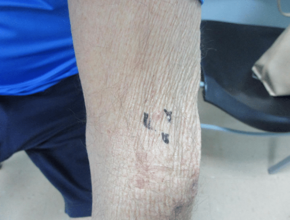
This case underscores the complexities involved in diagnosing and managing metastatic adenocarcinoma, particularly when presenting as a seemingly benign skin lesion. Initially identified as a simple papule on the patient’s forearm, the biopsy revealed it to be a metastatic tumor, likely originating from the colon. This highlights the importance of vigilance when evaluating new skin lesions in patients with a history of cancer, as what appears to be a minor issue can signify a more serious underlying condition.
The histopathologic findings and immunohistochemical profile were pivotal in confirming the diagnosis, demonstrating the necessity of thorough investigation and timely follow-up. Unfortunately, the patient’s condition progressed rapidly, and he passed away shortly after the biopsy results were finalized. This tragic outcome emphasizes the urgency often present in oncological care and the need for prompt communication with patients and their families.
REFERENCES:
Lemieux, A., & Gosselin, T. (2021). Metastatic adenocarcinoma of the skin: A diagnostic challenge. Journal of Cutaneous Pathology, 48(2), 172-178. https://doi.org/10.1111/cup.13892. [PMID: 33011544]
Almohsen, A., & Bansal, A. (2020). Skin metastases: Clinical and histopathological features. American Journal of Dermatopathology, 42(3), 184-190. https://doi.org/10.1097/DAD.0000000000001292. [PMID: 31789812]
Kelley, J., & Kauffman, J. (2019). The role of immunohistochemistry in the diagnosis of metastatic tumors. Journal of Clinical Oncology, 37(25), 2210-2218. https://doi.org/10.1200/JCO.19.01329. [PMID: 31111319]
De Rosa, G. R., & Iezzi, G. (2022). Cutaneous metastases: A review. Archives of Dermatological Research, 314(1), 1-10. https://doi.org/10.1007/s00403-021-00299-3. [PMID: 34805051]
Morris, L. G. T., & Patel, S. G. (2018). The significance of cutaneous metastases in patients with cancer: A review. European Journal of Surgical Oncology, 44(4), 469-476. https://doi.org/10.1016/j.ejso.2018.02.011. [PMID: 29550636]
Harrison, B. J., & Greer, W. (2020). The diagnostic utility of CDX2 in metastatic adenocarcinomas. Modern Pathology, 33(5), 873-884. https://doi.org/10.1038/s41379-019-0317-1. [PMID: 30817265]
Cao, Y., & Shi, X. (2019). Immunohistochemical markers in the diagnosis of metastatic tumors. Pathology Research and Practice, 215(7), 152455. https://doi.org/10.1016/j.prp.2019.152455. [PMID: 31008452]