CORRECT DIAGNOSIS:
Glomus Tumor
DISCUSSION:
Glomus tumors are benign neoplasms that originate from the cells of the glomus body, specifically from the arterial portion known as the Sucquet-Hoyer canal. This canal facilitates blood shunting between arterioles and venules, playing a crucial role in thermoregulation. Glomus bodies, composed of modified perivascular smooth muscle cells, are sensitive to temperature changes—contracting in response to cold and dilating to maintain blood flow to the nail bed, particularly in the fingertips.
Glomus tumors are most commonly found in the hands, particularly in the subungual region. Small, solitary tumors are the most frequent type, and they predominantly affect women, particularly those between the ages of 30 and 50.
Some genetic factors are associated with glomus tumors. A familial variant linked to chromosome 1p21-22 encodes the glomulin gene (GLMN) and presents as an autosomal dominant trait. Multiple glomus tumors may appear, often on the legs, and can occur in younger children. Additionally, patients with Neurofibromatosis type 1 (NF1) may develop multiple glomus tumors in the digits.
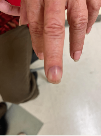
Clinically, glomus tumors are often painless but may present with a classic triad of symptoms: pain, pinpoint tenderness, and cold sensitivity. On physical examination, a small reddish or bluish papule several millimeters in diameter is typically noted. In some cases, they may present as longitudinal erythronychia with distal notching or overlying longitudinal fissures.
Diagnosis of glomus tumors can be supported by several clinical tests. One such test is Love’s Test, which involves using a sharp needle on the nail plate over the tumor; this application of pressure typically elicits pain in the affected region, indicating the presence of a glomus tumor. Another important diagnostic tool is the Hildreth Test, where a tourniquet is placed at the base of the digit, potentially alleviating the pain. Additionally, inflating a blood pressure cuff to 300 mmHg before or immediately after trauma can effectively abolish the pain response, further supporting the diagnosis. Lastly, assessing cold sensitivity is also helpful, as patients often experience exacerbated pain when an ice cube is placed on the nail or when the digit is submerged in cold water. These diagnostic tests are crucial for accurately identifying glomus tumors and distinguishing them from other conditions.
TREATMENT:
The primary treatment for symptomatic glomus tumors is surgical excision, which is aimed at relieving pain and removing the tumor entirely. Although these tumors are benign, there is a recurrence rate of up to 20%, making careful surgical technique and follow-up essential. Surgical excision typically involves removing the tumor along with a margin of surrounding tissue to minimize the risk of recurrence.
Given the location of the tumor and the patient’s persistent symptoms, referral to a hand surgeon is often necessary for optimal management, as they possess specialized expertise in the intricacies of hand anatomy and function. Our patient opted for this referral to a hand surgeon to ensure comprehensive evaluation and treatment, which may include preoperative imaging to assess the tumor’s size and depth and to plan the surgical approach. Postoperative follow-up will be crucial to monitor for any signs of recurrence and to evaluate the patient’s recovery.
CONCLUSION:
Glomus tumors are benign neoplasms originating from the glomus body, particularly the Sucquet-Hoyer canal, which is essential for thermoregulation. Commonly found in the hands, especially the subungual region, these tumors predominantly affect women aged 30 to 50. Genetic factors, including a familial variant linked to the glomulin gene (GLMN) and associations with Neurofibromatosis type 1 (NF1), may contribute to their development.
Clinically, glomus tumors may be painless but often present with pain, pinpoint tenderness, and cold sensitivity. Diagnosis is supported by tests such as Love’s Test, the Hildreth Test, and cold sensitivity assessments. Effective management involves surgical excision, with referral to a hand surgeon often recommended to ensure optimal care. Our patient’s choice to seek this referral emphasizes the importance of specialized treatment, with ongoing follow-up critical for monitoring recovery and preventing recurrence.
REFERENCES:
Lee, W., Kwon, SB., Cho, SH., Eo, SR., & Kwon, C. Glomus Tumor of the Hand. Archives of Plastic Surgery. 2015 May; 42(3): 295-301. [PMID 26015884]
Patel, T., Meena, V., & Meena, P. Hand and Foot Glomus Tumors: Significance of MRI Diagnosis Followed by Histopathological Assessment. Cureus. 2022 Oct; 14(10). [PMID 36381782]
Hazani, R., Houle, JM., Kasdan, ML., & Wilhelmi, BJ. Glomus tumors of the hand. Eplasty. 2008 Oct; 8:e48. [PMID: 18997858]
Yanai, K., Tajika, T., Kuboi, T., Hatori, Y., Umeyama, T., Arisawa, S., Katayama, A., Sano, T., & Chikuda, H. A case of solitary digital glomus tumor associated with neurofibromatosis type 1. SAGE open medical case reports. 2023 Aug; 11:2050313X231193984. [PMID 37609106]
Rosen, T., & Delman, H. (2021). Glomus tumors: Clinical features and management. Journal of the American Academy of Dermatology, 84(5), 1369-1374. https://doi.org/10.1016/j.jaad.2020.10.021. [PMID: 33184925]
Huang, J., & Zhang, Q. (2018). Familial glomus tumors: A case report and literature review. BMC Dermatology, 18(1), 6. https://doi.org/10.1186/s12895-018-0227-0. [PMID: 29599996]